
AI tumor volume estimations could improve prostate cancer treatment strategies
Artificial intelligence-estimated tumor volume may have more prognostic value than standard risk stratification methods, new research suggests.
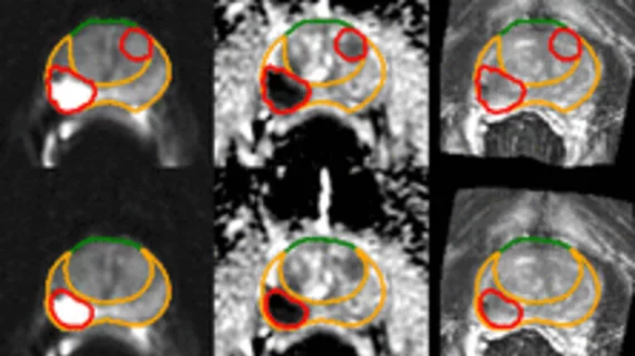
Researchers at Mass General Brigham recently developed an AI model that uses prostate MRI images to estimate total tumor volume. Following the model’s validation, it was retrospectively tested on a group of more than 700 prostate MRI exams to determine whether it could provide more consistent, detailed estimations of lesion size compared to human readers. Doing so could give providers more precise insight into how to best manage a patient’s treatment plan.
The group detailed the development and testing of their model Tuesday in the journal Radiology.
“Al-determined tumor volume has the potential to advance precision medicine for patients with prostate cancer by improving our ability to understand the aggressiveness of a patient's cancer and therefore recommend the most optimal treatment,” explained first author and founding member of the Mass General Brigham healthcare system David D. Yang, MD, of the Department of Radiation Oncology at Brigham and Women’s Hospital.
The team sought to determine whether the model’s size estimations, which indicate tumors’ aggressiveness, were associated with the patients’ outcomes 5 and 10 years after completing treatment—radical prostatectomy, radiation therapy, or both.
The segmentation algorithm was able to identify, accurately measure and outline around 85% of the most aggressive lesions, those that were ultimately given the highest PI-RADS scores by human readers. The team observed associations between the lesions with the highest AI tumor volume estimations and instances of cancer recurrence and metastases.
“The AI measurement itself can tell us something additional in terms of patient outcomes,” the group noted. “For patients, this can really tell them something about what are the chances of cure, and the likelihood that their cancer will reoccur or metastasize in the future.”
The researchers suggested that the algorithm's estimations could also make predicting a tumor’s aggressiveness much less time-consuming and cumbersome, potentially paving the way for initiating treatment earlier after a diagnosis.
Future research on the model will include larger sample sizes with different disease characteristics to ensure its generalizability.
The study abstract is available here.

In addition to her background in journalism, Hannah also has patient-facing experience in clinical settings, having spent more than 12 years working as a registered rad tech. She began covering the medical imaging industry for Innovate Healthcare in 2021.