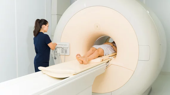
MRI could be 'powerful tool' for guiding surgery decisions in patients with rectal cancer
Specific image findings on MRI exams could prevent patients with rectal cancer from having to undergo surgery.
A recent analysis in the journal Radiology details findings that are related to the odds of survival and recurrence in patients who have undergone initial treatment for rectal cancer. While the utility of MRI in managing rectal cancer treatment has not been well studied thus far, experts involved in this new research suggest that their findings relative to the modality are promising.
“After undergoing chemotherapy and radiation for rectal cancer, patients are understandably concerned whether their cancer is gone or whether there may be some leftover disease. Using newer MRI techniques, we are now able to predict much better than in the past whether any cancer remains and, if so, whether it will come back and spread,” Krishnaraj, MD, a radiologist and director of UVA Health’s Division of Body Imaging, said in a release.
Most rectal cancers are initially treated with chemotherapy and radiation. However, for many, treatment can require surgery to remove a significant portion of the bowel, leaving patients with lifelong adjustments, including the use of a colostomy bag.
Researchers sought to determine if certain MRI findings in this population were indicative of recurrence and survival. If so, such information could be used to guide decision-making in how to further manage patients’ disease, potentially sparing some of invasive and consequential surgery.
For the study, the team examined the cases of 277 patients included in the Organ Preservation in Rectal Adenocarcinoma (OPRA) trial. Each patient underwent restaging MRI around two months after their initial treatment and received follow-up care for an average of four years. Researchers used imaging to group patients into three categories of treatment response: clinical complete response (cCR), near-complete clinical response (nCR) or incomplete clinical response (iCR).
Nearly half of the patients showed residual disease on imaging. Five-year disease-free survival for participants with cCR, nCR, and iCR was 81.8%, 67.6%, and 49.6%. Patients with cCR had greater organ preservation in comparison to those with nCR. The team found that the presence of restricted diffusion and abnormal nodal morphologic features were both significantly associated with residual disease.
“No one wants to get surgery if they can avoid it,” Krishnaraj said. “Now we have a powerful tool to help patients and their doctors predict who would benefit from surgery after initial chemotherapy and radiation and who can likely avoid surgery.”
The team also suggested that additional diagnostic tools, such as post-treatment endoscopies, could be used in conjunction with MRI to provide even greater insight into patients’ need for surgery. This is the focus of the group’s ongoing research.
“I am optimistic that continued advancement in MRI and other tools like endoscopy will provide better information about future outcomes,” Krishnaraj said. “Ultimately, I would love to get close to 99% predictive probability in better informing our patients about their potential risk for recurrence or spread of their cancers following treatment.”
The study abstract can be found here.

