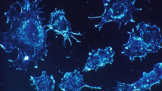
Microwave imaging method may improve cancer screening, treatment monitoring
A new molecular imaging method developed by engineers from Lehigh University in Bethlehem, Pennsylvania, may improve cancer screening and treatment monitoring through high-frequency microwaves.
The lab-on-a-chip method—which characterizes the nucleus of a single cell that is captured, analyzed and released by a small microfluidic device and penetrated with microwave technology—would be portable, faster and more cost efficient than current methods, according to an Aug. 14 Lehigh University release.
“High-frequency microwave has the advantage of being able to penetrate through the cell and into the nucleus, like an x-ray is able to do for the human body, but without harming the cell,” said Xuanhong Cheng, PhD, an associate professor of materials science and engineering at Lehigh University, in a prepared statement.
Together with James Hwang, PhD, professor of electrical engineering and computer science at Lehigh, Cheng plans to create the microwave equivalent of optical coherence tomography to see the inside of a cell nucleus and observe changes in nuclear morphology and DNA content. The goal is to better understand cell development and cancer progression, according to the news release.
Their goal is to ultimately advance their hardware to achieve greater detection sensitive and advance their software to create more detailed computer models. The research was funded by a three-year grant awarded by the National Science Foundation.

