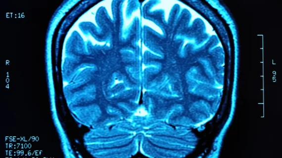
Unused MRI information offers insights into brain conditions
MRI scans produce mounds of unused data. Now, researchers at Washington University School of Medicine in St. Louis have created a new method of utilizing that information to determine how many and where cells have been lost due to injury or disease.
“There’s no easy way to detect the loss of neurons in living people, but such loss plays a role in many neurological diseases,” said Dmitriy Yablonskiy, PhD, and professor of radiology at Mallinckrodt Institute of Radiology, in a news release. “We’ve developed a method that takes a six-minute scan and tells you what types of cells are there and how extensively they’re connected.”
Yablonskiy and colleagues looked at background data from MRIs and discovered a signal that remained steady throughout the brain while a participant performed a certain task. These signals— labeled R2t*—varied across the brain.
After comparing R2t* data to that in the Allen Human Brain Atlas—which maps where genes are active throughout the brain— the group found three sets of gene networks that corresponded to R2t* signals. Activity in those genes intensified where the signal was strongest, and vice versa. Yablonskiy et al. concluded these groups of genes reflected the varying types and number of brain cells, along with their interconnectivity.
The team tested their method on the hippocampus of patients with Alzheimer’s disease and found those regions to be smaller in participants with AD compared to healthy people. Additionally, the remaining portion had lost cells and was in the process of decaying.
“There are MRI scans now that can detect brain atrophy even before people show symptoms of Alzheimer’s disease,” Yablonskiy said. “Our technique can show the brain degrading even before it begins to atrophy."
They are currently working to extend their technique to better understand brain diseases and disorders such as Alzheimer’s disease, schizophrenia, multiple sclerosis and more. They also seek to illuminate the development and growth of healthy brains, according to the release.
Research was published online Sept. 24 in Proceedings of the National Academy of Sciences.

