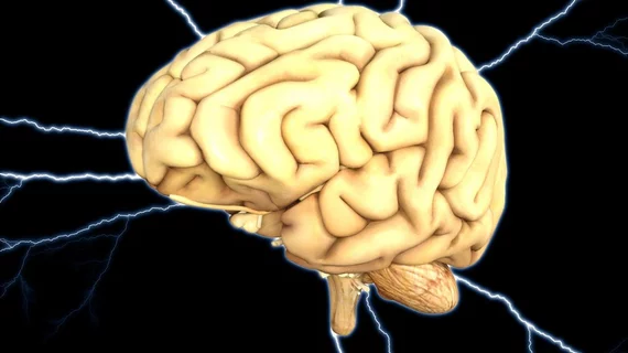
MRI reveals the bigger the brain, the greater the cancer risk
Researchers from the Norwegian University of Science and Technology (NTNU) in Trondheim, Norway have found that having a large brain—or greater intracranial volume—increases the risk for brain cancer.
"Several studies have shown that the size of different organs is an important factor in cancer development. For example, women with larger breasts have a greater risk of breast cancer. We wanted to check if this was also the case for brain tumors," lead author Even Hovig Fyllingen, a PhD candidate at NTNU, said in a prepared statement.
For the study, Fyllingen and colleagues used data from the Nord-Trøndelag Health Study (HUNT), which is comprised of health information and blood samples from thousands of Norwegians in the Nord-Trøndelag county region. The data was compared to a neurosurgery database at St. Olavs Hospital in Trondheim, Norway.
Data on those who had surgical interventions for high-grade brain tumors between 2007 and 2015 from both cohorts were compared to healthy controls from the HUNT study.
Intracranial volume was calculated from pretreatment three-dimensional (3D) T1-weighted MRI brain scans from 124 patients with high-grade glioma and 995 general population–based controls, according to the researchers.
Overall, they found brain tumors were more associated with a higher intracranial volume and were more prevalent in women than men.
“Intracranial volume is strongly associated with risk of high-grade glioma. After correcting for intracranial volume, risk of high-grade glioma was higher in women,” the researchers wrote. “The development of glioma is correlated to brain size and may to a large extent be a stochastic event related to the number of cells at risk.”
The research was published online in the August issue of the Oxford Academic journal Neuro-Oncology.

