Neuroimaging detects severe abnormalities from Zika virus in 15% of infants
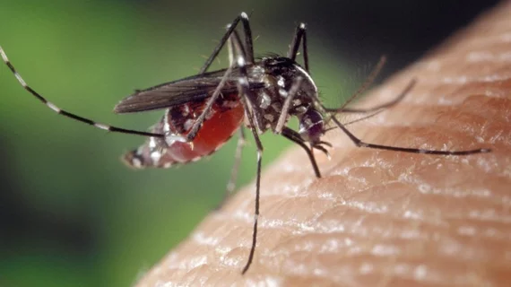
The Zika virus epidemic in Rio de Janeiro, Brazil from September 2015 to June 2016 began as a relatively unknown mosquito-borne virus which was later linked to microcephaly and other neurological abnormalities in a startling number infants in South and North America and other countries.
However, a new study has found that brain imaging with MRI, CT or ultrasound may predict neurodevelopmental health in infants who have been exposed to the virus in utero, according to research published online Dec. 12 in the New England Journal of Medicine.
For the study, Karin Nielsen, MD, professor of clinical pediatrics in the Division of Infectious Diseases at the University of California, Los Angeles (UCLA) Children's Hospital, and colleagues conducted a series of exams on 131 children who has been exposed to the Zika virus while in utero.
The exams included one imaging exam (using transfontanelle cerebral ultrasound, CT or MRI), an eye exam, a standard screening called Bayley III (measures infants’ cognitive, language and motor skills) and a brainstem evoked audiometry test which diagnoses hearing loss in newborns and young children.
By the age of 12 to 18 moths, 14.5 percent of children had at least one of three abnormalities. Specifically, significant problems were detected in seven of the 112 children (6.25 percent) who were evaluated for eye abnormalities; in six of the 49 children (12.2 percent) evaluated for hearing problems; and in 11 of the 94 children (11.7 percent) evaluated for severe delays in language, motor skills and/or cognitive function who also had brain imaging.
Neuroimaging, however, failed to predict severe neurodevelopmental delays in two percent of cases. Additionally, 16 percent of infants who had abnormal brain images early in life had normal cognitive, language and motor skills by 12 to 18 months of age, according to the researchers.
The study was funded by the National Institutes of Health (NIH)’s National Institute of Allergy and Infectious Diseases and National Eye Institute, the Thrasher Research Fund, the Bill and Melinda Gates Foundation Grand Challenges Explorations and government agencies and funders in Brazil.

