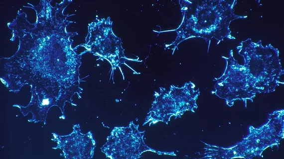
Radiotracer identifies 28 different forms of cancer
A team of German researchers has found a new class of radiopharmaceuticals capable of identifying 28 types of malignant tumors, imaging them with high uptake and image contrast.
The new radiotracer—68Ga-FAPI—attacks cancer associated fibroblasts, sometimes responsible for up to 90% of a tumor’s mass, co-author Uwe Haberkorn, MD, professor of nuclear medicine at the University Hospital of Heidelberg in Germany, and colleagues explained.
“Cancer associated fibroblasts have been described as immunosuppressive and as conferring resistance to chemotherapy, which makes them attractive targets for combination therapies,” Haberkorn added in a news release from the Society of Nuclear Medicine and Molecular Imaging (SNMMI).
Haberkorn et al. used PET/CT to scan 80 patients with 28 varying types of cancer. They set out to quantify 68Ga-FAPI uptake in primary, metastatic or recurring cancers. Each patient was facing an “un unmet challenge that could not be solved sufficiently with standard methods” and was referred for experimental diagnostics by their oncologist, according to the SNMMI release.
Overall, patients responded well to their exam. Mean SUV, median and range of 68Ga-FAPI in primary tumors and metastatic lesions were all similar. The highest average SUVmax (defined as greater than 12) was noticed in sarcoma, esophageal, breast, cholangiocarcinoma and lung cancer. Lowest uptake (average SUVmax less than 6) was in pheochromocytoma, renal cell, differentiated thyroid, adenoid cystic and gastric cancers.
“The remarkably high uptake of 68Ga-FAPI makes it useful for many cancer types, especially in cases where traditional 18F-FDG PET/CT faces limitations,” said Haberkorn, in the same release. “For example, low-grade sarcomas generally have a low uptake of 18F-FDG, causing an overlap between benign and malignant lesions. In breast cancer, 18F-FDG PET/CT is commonly used in recurrence, but not generally recommended for initial staging. And for esophageal cancer, 18F-FDG PET/CT often has only a low to moderate sensitivity for lymph node staging.”
The full study was published in the Journal of Nuclear Medicine.

