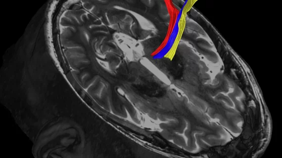
Refined MRI techniques pinpoint key treatment spot for Parkinson’s disease
A handful of new MRI methods can better target small portions of the brain tied to Parkinson’s disease and essential tremor and may result in safer, more effective treatments.
UT Southwestern Medical Center researchers in Dallas say these new imaging techniques allow neuroradiologists to analyze “pea-sized” areas in the brain’s thalamus. With these images, physicians can then burn away the damaged tissue using high-intensity focused ultrasound, the group wrote June 14 in Brain.
"The benefit for patients is that we will be better able to target the brain structures that we want," Bhavya R. Shah, MD, an assistant professor of radiology and neurological surgery at UT Southwestern's Brain Institute, said in a statement. "And because we're not hitting the wrong target, we'll have fewer adverse effects."
Those negative consequences—including problems walking or slurring words—are typically temporary, but become permanent in 15% to 20% of cases.
MRI-guided, high-intensity ultrasound has been used to destroy small areas of the thalamus linked to brain disorders for nearly a decade now, the authors noted. It’s also currently approved to treat essential tremor and tremors in patients with Parkinson’s, but has historically been unable to locate the “pea-sized” nucleus required to effectively combat such conditions.
Of the three MRI techniques described in the study, diffusion tractography is believed to be the most promising, Shah noted, due to its ability to record the natural water movements within tissues.
Quantitative susceptibility mapping—which picks up on distortions in the magnetic field caused by iron or blood—is also a viable approach. Fast gray matter acquisition TI inversion recovery—which illuminates both white and gray matter in further detail—was also effective.
The procedures have all been approved by the U.S. Food and Drug Administration for use in patients and UT Southwestern will begin testing them in its Neuro High Intensity Focused Ultrasound Program opening this fall. Shah and colleagues are also partnering with the Mayo Clinic on a multicenter clinical trial evaluating diffusion tractography in patients.

