Detection system outperforms radiologists at spotting chest pathologies on standard X-rays
A computer-aided detection tool outperformed radiologists at spotting difficult-to-diagnose chest pathologies, according to new data published in Academic Radiology.
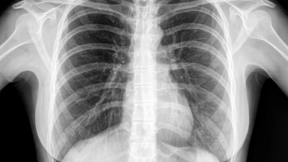
For the retrospective study, 197 patients underwent standard posteroanterior (PA) chest X-rays and a chest CT. Three blinded radiologists (two senior, one resident) were instructed to look for pulmonary nodules, infectious consolidation, pneumothorax, pleural effusion, aortic calcification, cardiomegaly and rib fractures among the images.
The radiologists analyzed dual-energy subtracted radiographs, while the CAD software reviewed standard PA chest X-rays. The CT scans were used as a reference.
The sensitivity of the CAD software proved to be significantly higher in the detection of infectious consolidation (67.9% versus 26.8%) and pulmonary nodules (54% vs. 35.6%). It did appear to perform slightly better in observing pneumothorax and rib fractures as well. As far as the other pathologies are concerned, no compelling differences were distinguished.
The results of the study show that CAD's ability to detect these pathologies could ultimately save radiologists time when sifting through thousands of images daily, and thus improve patient flow and care.
“The application of CAD systems could be useful in assisting the decision-making process of radiologists and reduc[ing] missed abnormalities on CXR,” Gioia Fischer, MD, with the Institute of Diagnostic and Interventional Radiology at University Hospital Zurich and University of Zurich, and co-authors noted. “Altogether, CAD systems seem to have their value as a second reader for certain pathologies, until now, however, it has not been conclusively clarified at which experience level the software offers the greatest benefit.”
You can read the full study in Academic Radiology.

