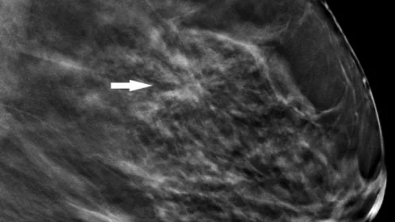
Research offers new guidance on managing architectural distortion visualized on DBT exams
Multiple areas of architectural distortion visualized on digital breast tomosynthesis (DBT) exams are likely to produce high-risk pathology results.
These new findings—shared this week in the American Journal of Roentgenology—could assist clinicians in decisions pertaining to the management of architectural distortion (AD), including how many suspicious areas merit invasive biopsy procedures.
Although the results of their work linked multiple ADs with high-risk pathology, the authors of the study shared that these findings did not necessarily increase malignancy rates.
“Multiple AD, compared with single AD, was significantly more likely to yield high-risk pathology, but was not significantly different in yield of malignancy,” corresponding author Lilian C. Wang, MD, and Northwestern Medicine co-authors explained. “In patients with multiple AD, multiple ipsilateral or contralateral AD commonly varied in pathologic classification (benign, high risk, or malignant).”
Since there is currently little guidance on the management of patients with multiple AD, Wang and colleagues conducted an analysis to compare outcomes of these patients to those who had just one AD identified on DBT. This included retrospectively examining the cases of 402 patients who underwent core needle biopsy of AD between April 2017 and April 2019.
Single AD was reported for 372 patients, and multiple AD for 30. The biopsy results for both groups were as follows:
Single AD: 145 benign, 121 high risk, 105 malignant, 1 other
Multiple AD: 10 benign, 35 high risk, 21 malignant
This translated into high-risk pathology rates of 52% in the multiple AD group and 32.5% in the single AD group. In comparison, malignancy rates did not vary as widely, at 31.8% vs 28.2%. The authors noted that the most aggressive pathologies varied between the two groups and were not found to be associated with the number of AD identified, prompting them to suggest that patients with multiple areas of architectural distortion may benefit from undergoing biopsy of all areas.
The study abstract can be viewed here.

