
Test bolus of diluted contrast optimizes scan timing during CTA
Using a double dose of diluted contrast for craniocervical CT angiography (cc-CTA) scans provides diagnostic quality imaging while also reducing the total amount of contrast required to do so.
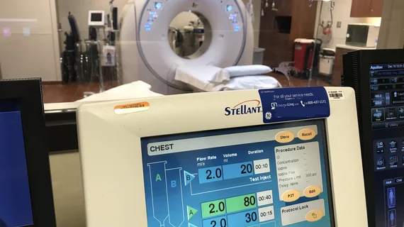
This is achieved by first giving patients a diluted dose via test bolus, prior to the timed examination. This enables techs to more precisely anticipate the CTA scan trigger time. Once that has been done, contrast containing less iodine is used for the primary CTA scan. During these scans, lowering the tube voltage and increasing the tube current provides quality visualization of vascular enhancement.
A new paper in Clinical Radiology details how the double low-dose protocol affects image quality while enhancing overall patient care.
“The success of CTA relies on optimal synchronization of CT scanning with [contrast media] passage. While the test bolus stands out as the most effective approach for anticipating the time delay in personalized CTA, the use of only a small dose of CM (usually 10 ml) results in considerable variation in arterial enhancement between patients, complicating the prediction of the actual enhancement of CTA,” Jun Li, with the Department of Radiology at Wujin Hospital Affiliated to Jiangsu University in China, and colleagues explained.
For the analysis, the team divided 86 consecutive patients undergoing cc-CTA into two groups—one (Group A) that received a standard dose of contrast (50 ml of 370 mgI/ml iopromide) at standard tube voltage and current, and one (Group B) that received a contrast with lower iodine content (270 mgI/ml iodixanol). Group B was also given a test bolus of 27 ml of diluted contrast.
For both groups, image quality was considered diagnostic. There were no significant differences observed between arterial densities of the ascending aorta or basilar artery between the groups, while there were significantly lower values of the common carotid artery, internal carotid artery, and middle cerebral artery in Group B.
What’s more, the effective dose and iodine uptake also were significantly lower in Group B.
The group suggested that these results could be due, in part, to the use of newer reconstruction algorithms, such as iterative reconstruction technology, deployed to reduce image noise. This allows for lower radiation doses without sacrificing image quality.
“Our study showed that when dual-source CT scanning with low-voltage and low-dose CM was applied to cc-CTA, the radiation dose was significantly reduced, while no significant reduction in the subjective scores was observed,” the authors noted. “These results indicate that combined with iterative reconstruction, appropriately cutting down the tube voltage can effectively reduce the radiation load, and images with diagnostic quality can also be obtained.”
The study abstract is available here.

In addition to her background in journalism, Hannah also has patient-facing experience in clinical settings, having spent more than 12 years working as a registered rad tech. She began covering the medical imaging industry for Innovate Healthcare in 2021.