Adaptable AI system detects subtle changes in imaging over time, has potential for multiple clinical settings
A new versatile artificial intelligence system can predict outcomes for a variety of pathologies based on assessments of longitudinal imaging datasets.
The Learning-based Inference of Longitudinal imAge Changes, or LILAC, system harnesses the power of machine learning to review medical images that have been collected over a prolonged period of time. LILAC was designed to offer greater training flexibility.
Unlike other AI models, it does not require extensive preprocessing or customization of data, as it can automatically detect subtle alterations and adjust accordingly—something that developers typically must do manually themselves. It does this by ignoring irrelevant details in images and instead highlighting the time-dependent details of interest.
“This new tool will allow us to detect and quantify clinically relevant changes over time in ways that weren't possible before, and its flexibility means that it can be applied off-the-shelf to virtually any longitudinal imaging dataset,” study senior author Mert Sabuncu, PhD, vice chair of research and a professor of electrical engineering in radiology at Weill Cornell Medicine, explained. “This enables LILAC to be useful not just across different imaging contexts but also in situations where you aren’t sure what kind of change to expect.”
The tool can be used in a variety of settings, including on images from pathology and radiologic datasets. It has been tested in multiple proof of concept studies ranging from imaging of tissue samples and pathology slides to MRI scans of the brain.
In the MRI assessment, LILAC was tasked with determining how much time had passed in between multiple series of brain scans a group of older adults had undergone. Researchers also tested its ability to predict cognitive scores based on the imaging from a group of adults with mild cognitive impairment. In both cases, LILAC outperformed standard methods of evaluation by 40%. In this sense, it could be used to predict who may be susceptible to cognitive decline in the future.
It also was tested on microscope images showing in-vitro-fertilized embryos as they develop, in addition to a group of images from new embryos. For this analysis, LILAC was prompted to determine which images had been taken earlier. And in a similar experiment, it was presented with images of a wound taken over time and tasked with placing them in chronological order based on signs of healing tissue. In both tests, LILAC achieved 99% accuracy.
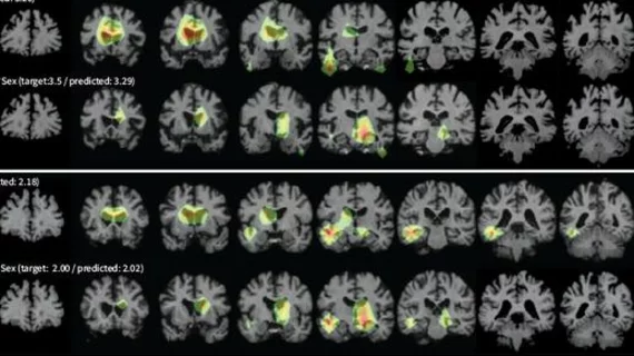
As the reliability of AI often comes into question, the team took their work a step further by training LILAC to highlight the imaging features that were most relevant to its predictions. This further enhances its applicability to a wide range of clinical uses, the group suggested.
“LILAC provides a streamlined approach that allows for easy adaptation to various types of longitudinal image datasets, minimizing the complexity and customization often associated with other methods,” they noted. “This flexibility makes LILAC accessible to a wide range of applications, while its simplicity ensures that it can be implemented without the need for extensive customization.”
Learn more about the findings here.

