A recent analysis found that a significant amount of studies do not provide information pertaining to their raw data, source code or model. As a result, up to 97% of these studies do not produce systems that are fit to be used in real-world clinical scenarios.
Prior to 2018, it was recommended that women at higher-than-average risk undergo supplemental breast MRI screening only if they had a personal history of breast cancer in addition to carrying a hereditary breast cancer gene mutation.
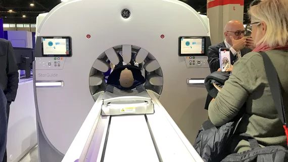
Example of a new CZT digital detector technology SPECT system, the GE Starguide SPECT-CT system on display at RSNA 2022. The system uses arms that extend to the patient so the detectors are closer to the photon emission source inside the patient to improve image quality.
Washington University is using a novel low-count quantitative SPECT technique to measure the concentration of alpha particle radiopharmaceutical therapy activity in the tumor and in radio-sensitive organs.
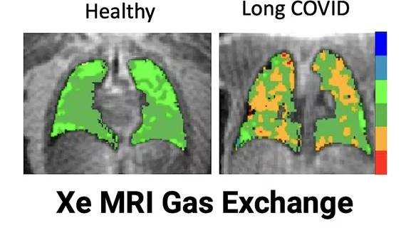
Sean Fain, PhD, vice chair of radiology and research and a professor of radiology, University of Iowa, discusses how long-COVID lung damage can be tracked using xenon (Xe) gas MRI and quantitative CT at RSNA 2022.
AI-automated detection of an intracranial hemorrhage on CT. The software can alert the care team before a radiologist even sees the exam. Example shown by TeraRecon at RSNA 2022.
Sanjay Parekh, PhD, senior market analyst with Signify Research, explains how some radiology AI is being adopted outside of radiology departments to improve care.
Study author Alexis M. Stranahan, PhD, is a neuroscientist at the Medical College of Georgia at Augusta University. Photo Credit: Michael Holahan, Augusta University
Researchers examined male and female mice on high-fat diets to learn about the impacts of fat distribution on brain inflammation.
Use of the template, which included PCP notifications, also resulted in an increase of biochemical testing, follow-up imaging and specialist referrals in patients with incidental adrenal masses.
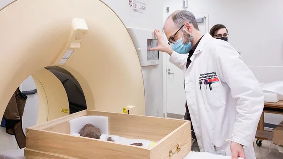
Nick Gruszauskas running the fossils through CT scanner. Credit: UChicago Medicine
Experts are hoping that, by imaging the dinosaur’s shoulder girdle and forelimb, they can create a digital model that could help in determining a T. Rex arm’s range of motion and strength.
Tim Szczykutowicz, PhD, DABR, associate professor of radiology at the University of Wisconsin-Madison, is helping develop a new type of photon-counting CT detector that was shown as a work-in-progress by GE Healthcare at RSNA 2022.