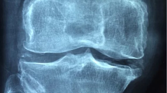
3D mapping algorithm reads knee MRIs for new arthritis treatments
A new algorithm developed by researchers at the University of Cambridge can create a 3D model based on a patient’s knee MRI to help clinicians develop novel treatments for joint arthritis.
The technique, known as 3D cartilage surface mapping, detects subtle changes in a person’s knee joint that cannot be picked up via conventional x-ray or MRI. This information will be a “considerable boost” to creating new treatments for osteoarthritis, experts wrote recently in the Journal of Magnetic Resonance Imaging.
"We don't have a good way of detecting these tiny changes in the joint over time in order to see if treatments are having any effect," James MacKay from Cambridge's Department of Radiology, said in a news release. “In addition, if we're able to detect the early signs of cartilage breakdown in joints, it will help us understand the disease better, which could lead to new treatments for this painful condition."
The team tested their approach on knee joints taken from bodies donated for medical research and another 14 individuals between 40 and 60 years old. Each participant suffered from mild to moderate knee OA, but was deemed too young for a knee replacement.
After comparing joints in those with the knee arthritis to healthy individuals, MacKay and colleagues found their approach could accurately monitor small changes over a six month period.
"There's a certain degree of deterioration of the joint that happens as a normal part of aging, but we wanted to make sure that the changes we were detecting were caused by arthritis," MacKay added. "The increased sensitivity that 3D-CaSM provides allows us to make this distinction, which we hope will make it a valuable tool for testing the effectiveness of new therapies."
The software is available for free and can be integrated into existing systems, and training radiologists on how to use the algorithm in their daily workflow is “short and straightforward,” the group added.

