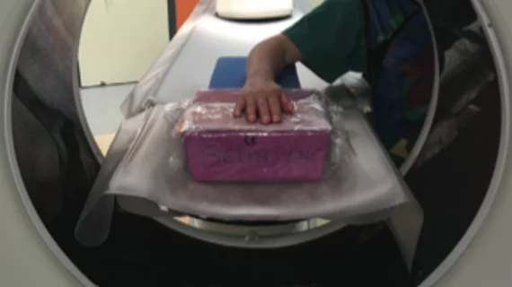
4D CT could supplement wrist radiographs in evaluating radioulnar deviation
For patients with abnormal wrist motion, four-dimensional (4D) CT imaging during radioulnar deviation could improve diagnosis of suspected scapholunate ligament tears and injuries.
The findings published online Sept. 25 in Radiology may limit the need for patients to undergo more invasive imaging procedures such as CT arthrography, MR arthrography and arthroscopy.
The researchers also noted that 4D CT is accurate enough to serve as a supplementary tool in evaluating patients with inconclusive radiographs.
Aymeric Rauch, MD, from the Guilloz Imaging Department at the University Hospital Center of Nancy in France, and colleagues analyzed the wrists of 37 participants at an average age of 38 years. All were suspected of having scapholunate instability and were examined with 4D CT and CT arthrography. An additional 23 controls were included for analysis.
“Five angular parameters for radioscaphoid angle (RSA) and lunocapitate angle (LCA) variation during radioulnar deviation were calculated by two independent readers,” the researchers wrote. “CT arthrography was used as the reference standard method for scapholunate ligament tear identification.”
Study results included the following:
In the control group, the mean values for RSA were 103° and 104° and the mean values for LCA were 86° and 90° with a coefficient of variation of 11 percent, and 13 percent for reader 1 and reader 2.
The interobserver and intraobserver agreements were "excellent" for RSA and substantial to excellent for LCA, researchers wrote.
In the pathologic group (n = 14), LCA amplitude, standard deviation, and maximal angle were lower for both readers with respect to the control group, measuring 36 percent and 44 percent, 37 percent and 44 percent, and 13 percent and 19 percent.
RSA amplitude did not show statistically significant results in the pathologic group. LCA yielded the highest sensitivity (71 to 93 percent), whereas RSA yielded the highest specificity (87 to100 percent).

