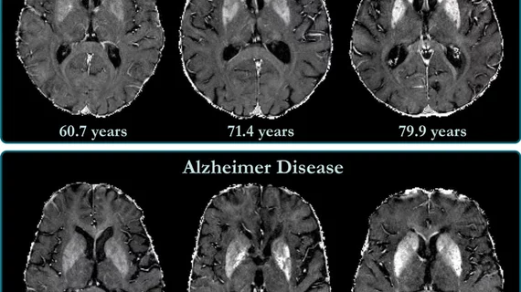
Brain iron buildup is associated with cognitive decline in Alzheimer’s patients
Iron accumulation in the exterior layer of the brain shown on MRI is associated with cognitive deterioration in patients with Alzheimer’s disease, according to a new study published Tuesday.
Researchers from the Medical University of Graz in Austria compared baseline levels of brain iron in patients with the memory disease and those without it. Individuals in the former group contained higher amounts in multiple regions of their brains, including areas involved in language and consciousness, the group reported in Radiology.
“We found indications of higher iron deposition in the deep gray matter and total neocortex, and regionally in temporal and occipital lobes, in Alzheimer’s disease patients compared with age-matched healthy individuals,” Reinhold Schmidt, MD, chairman of the department of neurology at Graz, said June 30. He added that these findings may push treatment approaches toward drugs that reduce excess iron from the body.
Iron deposition has previously been tied to amyloid beta, a protein that accumulates in the brains of those with Alzheimer’s and inhibits cellular function. There has been little insight into how this unfolds in the neocortex, however, due to challenges with imaging this outer brain layer.
To overcome these issues, Schmidt et al. developed a new 3T MRI approach, coupled with postprocessing, to eliminate image distortions. They scanned 100 patients with AD and 100 healthy controls who were, on average, 73 years old. Out of the entire Alzheimer’s group, 56 individuals underwent neuropsychological testing and brain MRI follow-up at 17 months, on average.
The researchers concluded that iron buildup was associated with cognitive decline independently of brain volume loss. And fluctuations in iron levels over time in the temporal lobe were also linked to patients with AD.
Efforts to treat or even slow the progression of Alzheimer’s have largely fallen flat, the group noted. But drugs that eliminate excess iron, known as chelators, should be a logical next step.
“Our study provides support for the hypothesis of impaired iron homeostasis in Alzheimer’s disease and indicates that the use of iron chelators in clinical trials might be a promising treatment target,” Schmidt said. “MRI-based iron mapping could be used as a biomarker for Alzheimer’s disease prediction and as a tool to monitor treatment response in therapeutic studies.”

