
CTA-based radiomics can reliably estimate time since stroke onset
The use of radiomics features can help clinicians estimate time since stroke onset based on CTA images.
Time since stroke onset (TSS) is imperative in the management of patients who have suffered an acute ischemic stroke (AIS). Patients who are treated via thrombolysis within a 4.5-hour window of the onset of AIS have the best outcomes, but determining TSS can be challenging, authors of a new paper published in Academic Radiology explained.
“The accurate assessment of TSS is essential for stroke treatment and prognosis. Nonetheless, TSS is determined only in a few cases, and many patients do not receive correct and timely treatment,” corresponding author Lei Zhang, MD, PhD, with the Department of Radiology at Jiangnan University Medical Center, and colleagues noted.
MRI is a valuable tool for estimating TSS, but due to access issues and patient contraindications, its use is limited. CT angiography (CTA) is often used as an alternative to MRI, but depending on when the exam is acquired in AIS patients, it also has limited utility in TSS determination.
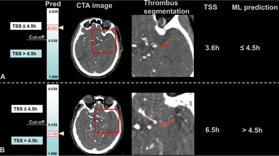
In recent years, the use of radiomics features in these sorts of scenarios has proven beneficial in predicting outcomes, determining malignancy, etc. That’s what led experts involved in this most recent research to hypothesize that CTA-based thrombus radiomics could help in identifying patients who are within a crucial treatment window following AIS.
The retrospective analysis included 221 patients with AIS. Each patient had undergone CTA, from which more than 900 radiomics features were extracted to construct a predictive machine learning model. Experts also developed a model that included clinical features alongside the radiomics features to determine whether they would improve the model’s predictive value.
Upon comparison, researchers found that both models performed well in TSS estimation, achieving similar (radiomics model alone: .803, combined model: .813) AUCs in both training and testing datasets.
Among the radiomics features selected, the two that reflected the distribution of voxel intensities in the image and differences in grayscale intensity in the thrombus were found to be the most effective.
“The composition of a thrombus changes over time and is shown as changes in grayscale intensity and textural characteristics on radiological images,” the authors explained.
The authors suggested that radiomics features could be especially useful for determining TSS in scenarios where clinical data is lacking, such as when patients have altered mental status. They added that this could trigger more prompt treatment by identifying acute ischemic stroke patients within a specific treatment window.
The study abstract is available here.

In addition to her background in journalism, Hannah also has patient-facing experience in clinical settings, having spent more than 12 years working as a registered rad tech. She began covering the medical imaging industry for Innovate Healthcare in 2021.