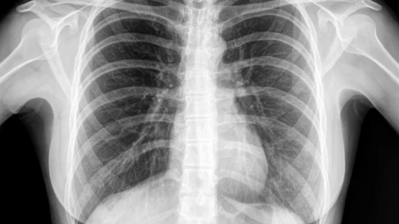
Deep learning bests radiologists at classifying thoracic disease on chest x-rays
A deep learning algorithm consistently outperformed radiologists at classifying chest x-rays with thoracic diseases, according to a recent study published in JAMA Network Open.
The deep learning-based automated detection algorithm (DLAD) was trained using more than 53,000 chest x-rays with normal findings and more than 34,000 abnormal chest x-rays showing one identified major thoracic disease, which included pulmonary malignant neoplasms, active pulmonary tuberculosis, pneumonia and pneumothorax.
The DLAD notched a median area under the ROC curve score of 0.979 for image-wise classification and 0.972 for lesion-wise localization. When compared to three physician groups—non-radiologist physicians, radiologists and thoracic radiologists—the algorithm proved superior at both tasks.
Importantly, the algorithm was externally validated on 486 normal chest radiographs and 529 abnormal chest radiographs taken from four different institutions across various continents. It was also validated using in-house images, including 300 normal chest x-rays and 789 abnormal chest x-rays.
“This consistent performance of the DLAD across the external validation data sets acquired from different populations suggests that our DLAD’s performance may be generalized to various populations,” wrote Eui Jin Hwang, MD, department of radiology at Seoul National University College of Medicine in South Korea, and colleagues.
Hwang and colleagues also found “significant improvements” in readers’ image-wise classification (0.814-0.932 to 0.904-0.958 ROC) and lesion-wise localization ((0.781-0.907 to 0.873-0.938) when they used the DLAD as a second reader.
“The DLAD can contribute to reducing perceptual error of interpreting physicians by alerting them to the possibility of major thoracic diseases and visualizing the location of the abnormality,” the authors added. “In particular, the more obvious increment of performance in less-experienced physicians suggests that our DLAD can help improve the quality of CR interpretations in situations in which expert thoracic radiologists may not be available.”

