
Deep learning automatically measures key features of TBI
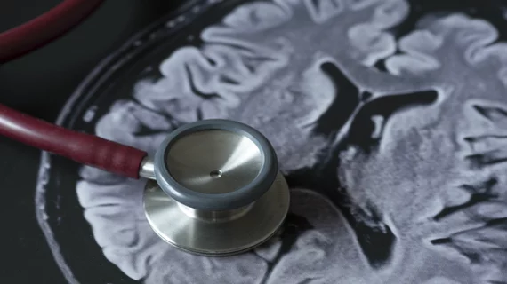
A team of international researchers has developed a deep learning system capable of reliably detecting and quantifying lesions associated with traumatic brain injury from patients’ CT scans.
The system, outlined May 14 in The Lancet: Digital Health, utilizes data from multiple institutions across Europe and was validated using scans from more than 500 patients in India. Compared with manual assessment, the CNN produced similarly accurate measurements, allowing clinicians to quantify lesion burden and progression.
First author Miguel Monteiro, with Imperial College London, and colleagues made their algorithm free for use and believe it has real-world clinical applications.
“The ability to automatically monitor lesion progression offers key opportunities to improve patient stratification, guide and monitor management, and investigate potential causes and risk factors for lesion progression in large cohort studies…,” the researchers noted. “Until now, the identification of factors that predict or cause contusion progression, or both, has been hampered by the need to estimate lesion volume and change manually, restricting analyses to small sample sizes.”
The researchers trained and validated their convolutional neural network on manual segmentations performed in 98 scans. That algorithm was then used to segment a new dataset of scans which comprised 839 CTs from 38 institutions. Their model accurately segmented, quantified, and detected multiclass hemorrhagic lesions and perilesional oedema.
Specifically, these measurements can aid in image-based diagnoses, determine brain injury type, quantify severity, and gauge injury progression.
The size and diversity of the data used in this research make the findings applicable to real-world clinical situations, the authors noted.
“Such algorithms will find clear research applications, and, if adequately validated, may be used to help facilitate radiology workflows by flagging scans that require urgent attention, aid reporting in resource-constrained environments, and detect pathoanatomically relevant features for prognostication and a better understanding of lesion progression,” Monteiro and colleagues added.
They acknowledged that their CNN was less accurate at segmenting small hemorrhagic lesions, but these are less clinically important in terms of determining prognosis or therapeutic approaches, the authors wrote. These lesions are typically microhemorrhages, they added, and their clinical significance is based on quantity not individual volumes.

Matt joined Chicago’s TriMed team in 2018 covering all areas of health imaging after two years reporting on the hospital field. He holds a bachelor’s in English from UIC, and enjoys a good cup of coffee and an interesting documentary.