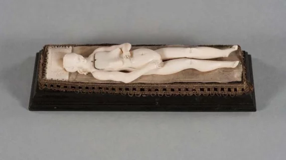
Historic medical objects imaged for insights
Duke researchers have used micro CT to peer deep into medicine’s past.
Specifically, the team imaged small ivory figurines used by German doctors in the late 1600s as they strived to understand and teach the workings of the human body.
The objects, called manikins, had removable models of organs and have long been conversation pieces among wealthy art collectors.
The researchers will present their new insights into the manikins at RSNA’s annual meeting next week in Chicago.
Fides Schwartz, MD, and colleagues chose micro CT for the modality’s ultrafine resolution and volumetric capabilities.
“This is potentially valuable to scientific, historic and artistic communities, as it would allow display and further study of these objects while protecting the fragile originals,” Schwartz says in a news release sent by RSNA. “Digitizing and 3D printing them will give visitors more access and opportunity to interact with the manikins and may also allow investigators to learn more about their history.”
Read the entire story below:

