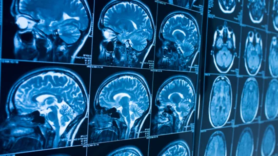
MRI reveals brain volume abnormalities in schizophrenia, other mental illnesses
Researchers from across the globe recently put their heads together to analyze mounds of neuroimaging data, and now have a better understanding of schizophrenia.
That’s according to a study incorporating hundreds of MRI scans, published Feb. 12 in the American Journal of Psychiatry. Upon analysis, patients with a rare chromosome disorder and known risk factors for schizophrenia (22q11DS), had lower brain volumes in certain regions of the organ. The findings may shed light on other serious mental illnesses, the researchers believe.
"We've pieced together many of the major research centers studying 22q11DS around the world to create the largest ever neuroimaging study of the disorder," first author Christopher Ching, PhD, a postdoctoral researcher at the University of Southern California’s school of medicine, said in a statement.
Individuals with 22q11DS deletion syndrome, or DiGeorge syndrome, are missing a small piece of chromosome 22, which can impact every all parts of the body. The abnormality is also the strongest known genetic risk factor for schizophrenia. In fact, nearly 25% of those with 22q develop the disease or experience psychotic symptoms, the authors noted.
Only about 1 in 4,000 individuals has this disorder, making it difficult for an institution to study it in isolation. But understanding the condition would offer insight into how psychiatric problems develop over specific time periods.
With this in mind, USC’s Enhancing Neuro Imaging Genetics through Meta-Analysis (ENIGMA) consortium created a working group to pool together 22q data from research centers across the world. In total, they looked at 533 MRI scans taken from patients with DiGeorge syndrome, along with 330 healthy control subjects.
Using a novel analytics methods created at the California university’s neuroimaging and informatics institute, the consortium measured and mapped structural variants between the brains of each group.
Overall, those with 22q showed “significantly” lower brain volumes, with lower measurements in the thalamus, hippocampus and amygdala, compared to controls. The former group also had higher volumes in some brain structures, the authors noted.
What’s more, brain changes in subjects with the genetic abnormality and psychosis “overlapped” with brain alterations reported in past neuroimaging-based studies of schizophrenia, bipolar disorder, major depression and obsessive compulsive disorder.
"That's important because these overlapping brain signatures add evidence to support 22q11DS as a good model for understanding schizophrenia in the wider population," he said. "And thanks to these large ENIGMA studies, we now have a way to directly compare standardized brain markers across major psychiatric illnesses on an unprecedented scale."

