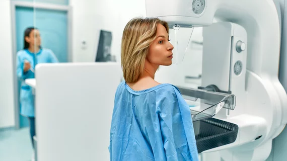
Screening mammography ‘signals’ predict women’s breast cancer risk
Separating women’s screening mammograms into those who are likely to develop breast cancer and patients at lower risk can reduce unnecessary imaging and associated costs. And Hawaiian researchers now say they have developed a tool capable of doing just that.
Experts from the University of Hawaii Cancer Center utilized more than 25,000 digital mammograms to develop their deep learning model. The tool searches for details, or signals, in scans that may indicate if patients are at an increased risk of cancer.
Testing showed the platform underperformed in assessing risk factors for interval cancers but beat out relying solely on clinical risk factors such as breast density. The findings highlight AI’s role as a second reader, particularly during screening exams, the team noted Sept. 7 in Radiology.
“The results showed that the extra signal we’re getting with AI provides a better risk estimate for screening-detected cancer,” said John A. Shepherd, PhD, a professor and researcher in the university’s Population Sciences in the Pacific Program. “It helped us accomplish our goal of classifying women into low risk or high risk of screening-detected breast cancer.”
More than 6,300 individual women who underwent screening mammography were included in the study. At least 1,600 developed breast cancer detected during screening, while 351 were diagnosed with interval invasive cancer.
Shepherd and co-authors believe their findings have important implications for practices using breast density alone to guide patient care. For example, a woman’s individual risk can determine if she needs to come in for another exam the following year or if she’s lower risk and may not require more frequent mammograms.
Furthermore, deep learning could inform decisions about adding supplemental MRI or other imaging for women in high-risk groups.
“By ranking mammograms in terms of the probability of seeing cancer in the image, AI is going to be a powerful second reading tool to help categorize mammograms,” Shepherd concluded.
Read the full study here.

