Artificial Intelligence

Artificial intelligence (AI) is becoming a crucial component of healthcare to help augment physicians and make them more efficient. In medical imaging, it is helping radiologists more efficiently manage PACS worklists, enable structured reporting, auto detect injuries and diseases, and to pull in relevant prior exams and patient data. In cardiology, AI is helping automate tasks and measurements on imaging and in reporting systems, guides novice echo users to improve imaging and accuracy, and can risk stratify patients. AI includes deep learning algorithms, machine learning, computer-aided detection (CAD) systems, and convolutional neural networks. 

Advanced viz global market to hit $2.8B by 2020

The advanced visualization market, including products for standalone workstations, enterprise-wide software and associated consulting and maintenance, has been projected for a compound annual growth rate of 10.9 percent from 2015 to 2020, reaching $2.8 billion by 2020, according to a new report published by MarketsandMarkets.

Thumbnail

Speeding up segmentation

Prior to extended hepatectomy, the tedious process of liver volume assessment must be conducted. Automated and semiautomated liver segmentation methods, however, stand to greatly increase the efficiency of this task, as evidenced by a recent study.

Thumbnail

How much does IR improve low-dose CT imaging quality? It depends on the scanner

Iterative reconstruction (IR) makes it possible to reduce radiation dosage without a reduction in imaging quality in low-contrast CT examinations, but its effectiveness varies with different doses and vendors, according to study results published online May 19 in the journal Radiology.

Patient motion during MRI proves to be costly conundrum

When a patient moves during an MRI, it can mean having to redo the scan. Repeated scans take time, and tied up scanners can affect a hospital’s bottom line. Just how costly is patient motion? A recent study from Harborview Medical Center in Seattle pegged the potential cost of motion artifacts at $115,000 per scanner per year.

Thumbnail

St. Louis surgical team uses 3D printing to plan toddler’s treatment

When cardiologists from Washington University in St. Louis were preparing to treat a toddler whose heart vessels were located in a position that made it tough for the child to breathe, they didn’t just rely on imaging. In another example of the power of 3D printing, the surgeons created a life-sized replica of the young boy’s heart.

Thumbnail

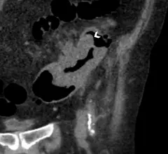
Dutch docs use CT colonography to distinguish carcinoma from diverticulosis

Features found at CT colonography have proven effective at discriminating between sigmoid cancer and diverticular disease, a discovery that could reduce unnecessary medical procedures and improve patient care, according to results of a study published in the April issue of Radiology.

Three babies breathing easier thanks to 3D printing

Time for a happy update on a story that appeared in Health Imaging and elsewhere a couple years ago.

Thumbnail

Video: Post-mortem CT paired with 3D printing helps determine cause of death

When the Victorian Institute of Forensic Medicine (VIFM) installed a CT scanner in their mortuary in 2005, it quickly became apparent how performing imaging exams on the dead could benefit the living.

Around the web

RBMA President Peter Moffatt discusses some of the biggest obstacles facing the specialty in the new year. 

Deepak Bhatt, MD, director of the Mount Sinai Fuster Heart Hospital and principal investigator of the TRANSFORM trial, explains an emerging technique for cardiac screening: combining coronary CT angiography with artificial intelligence for plaque analysis to create an approach similar to mammography.

A total of 16 cardiology practices from 12 states settled with the DOJ to resolve allegations they overbilled Medicare for imaging agents used to diagnose cardiovascular disease.