Artificial Intelligence

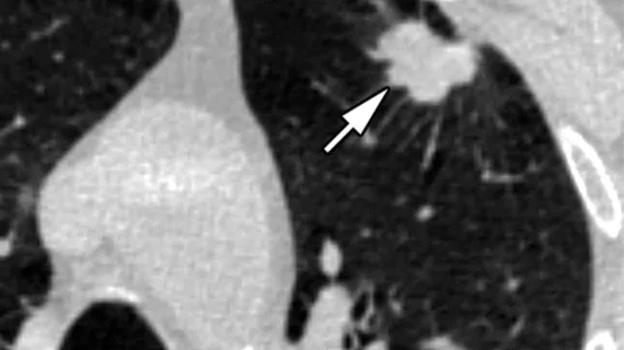
Artificial intelligence (AI) is becoming a crucial component of healthcare to help augment physicians and make them more efficient. In medical imaging, it is helping radiologists more efficiently manage PACS worklists, enable structured reporting, auto detect injuries and diseases, and to pull in relevant prior exams and patient data. In cardiology, AI is helping automate tasks and measurements on imaging and in reporting systems, guides novice echo users to improve imaging and accuracy, and can risk stratify patients. AI includes deep learning algorithms, machine learning, computer-aided detection (CAD) systems, and convolutional neural networks. 

Computer-extracted imaging features that ID prostate cancer: 5 findings

Using computer-extracted features to improve prostate cancer diagnosis at MR imaging has powerful potential, but what features are best to discriminate cancer from benign disease?

Thumbnail

Art meets medicine: New 3D tour of heart on display in Austrian museum

Medical images are not just profoundly useful at advancing care, they can also be quite beautiful. An example of art meeting medical research was recently put on display at the Ars Electronica Center in Linz, Austria.

Thumbnail

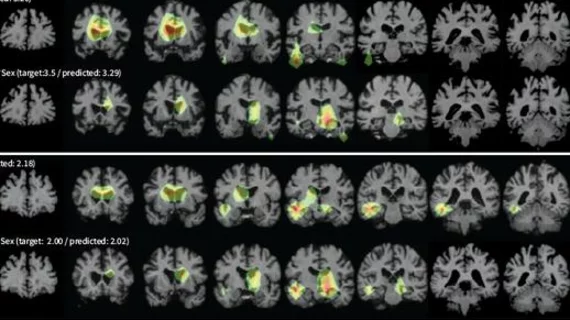
Heart-health risks show up in brain MRI as early predictors of Alzheimer’s

Drinking, smoking and chronic overeating are among the risky behaviors known to hurt not only the heart but also the brain. A new study combining cognitive testing with brain MRI provides a clearer picture of this gradual undoing of the mind in process.

Thumbnail

Plastic bot could offer assist during MR-guided surgery

Robot-assisted surgery and MR-guided procedures are two advances that are transforming medicine. The two are difficult to mix, however, given the strong magnetic fields of MRI scanners.

Cerner to Replace Department of Defense's Anatomic Pathology Laboratory Information System

The Defense Health Agency (DHA) has selected Cerner, which competed and successfully won an award, to replace the Military Health System's (MHS) existing anatomic pathology laboratory information system.

Thumbnail

Functional MRI shows brain changes after a single harrowing experience

First they thought they were about to die in a plane that had lost all power over the Atlantic Ocean. Then they survived an emergency landing on a chancy island airfield. Now some of the nearly 300 passengers of Air Transat Flight 236 are helping to show how post-traumatic stress disorder permanently alters the way the brain processes new information as well as memories—good, bad and neutral.

Researchers in Illinois putting spotlight on MR elastography

The University of Illinois and Carle Health System have begun work on testing MR elastography (MRE) for those with epilepsy and brain tumors.

Thumbnail

Two in one: Hybrid 3D printing integrates multiple modalities

Researchers have integrated CT with 3D transesophageal echocardiography (3DTEE) to create a 3D model of a patient’s heart, marking the first time multiple modalities have been combined to support 3D printing.

Around the web

RBMA President Peter Moffatt discusses some of the biggest obstacles facing the specialty in the new year. 

Deepak Bhatt, MD, director of the Mount Sinai Fuster Heart Hospital and principal investigator of the TRANSFORM trial, explains an emerging technique for cardiac screening: combining coronary CT angiography with artificial intelligence for plaque analysis to create an approach similar to mammography.

A total of 16 cardiology practices from 12 states settled with the DOJ to resolve allegations they overbilled Medicare for imaging agents used to diagnose cardiovascular disease.