Cardiac Imaging

While cardiac ultrasound is the widely used imaging modality for heart assessments, computed tomography (CT), magnetic resonance imaging (MRI) and nuclear imaging are also used and are often complimentary, each offering specific details about the heart other modalities cannot. For this reason the clinical question being asked often determines the imaging test that will be used.

Merit Medical Acquires Pressure-Assisted Hemostatic Devices From Datascope Corp.

Merit Medical Systems, Inc. (Nasdaq:MMSI), a leading manufacturer and marketer of proprietary disposable devices used primarily in cardiology, radiology and endoscopy, today announced that it has acquired from Datascope Corp. the SAFEGUARD(R) Pressure Assisted Device, which assists in obtaining and maintaining hemostasis after a femoral procedure, and the AIR-BAND(TM) Radial Compression Device, which is indicated to assist hemostasis of the radial artery puncture site while maintaining visibility.

T1 mapping allows for detection of deadly heart condition on MRI

A new MRI technique has been shown to be able to detect Fabry disease, a genetic condition that can result in fatal heart problems, according to a study published in the September issue of Circulation: Cardiovascular Imaging.

Facility comparison of NM exams support UltraSPECT as viable solution to meet ASNC guidelines

UltraSPECT, a leading provider of image reconstruction technology that reduces radiopharmaceutical dose and acquisition time for nuclear medicine (NM) exams, announces today that study data has shown UltraSPECT solutions are a viable method for meeting American Society of Nuclear Cardiology (ASNC) low dose guidelines. Furthermore, technologists are able to rely on their existing imaging protocols, providing added confidence in overall results. UltraSPECT will be demonstrating this solution to conference attendees at the upcoming ASNC Annual Meeting, September 26-29, in Chicago.“What this preliminary data shows us is that healthcare facilities do not have to change their standard imaging protocols to meet ASNC guidelines to lower dose,” explained Gordon DePuey, MD, director of Nuclear Medicine at St. Luke’s-Roosevelt Hospital, New York. “I am looking forward to seeing the great progress that the cardiovascular imaging community can make in 2014 as proven and viable solutions are applied to lower the dose for the benefit of patients and to continue to advance healthcare.”

MEDIAN Technologies Announces New Contract

MEDIAN Technologies (ALMDT), a leading medical imaging software solutions developer, and a service provider for image interpretation and management in oncology clinical trials, today announced that it has signed a €206K firm contract with one of its top 10 biopharmaceutical company client. The contract covers the implementation of an exploratory imaging biomarker, tumor volume, in a first-in-man phase I study on adult patients with advanced malignant solid tumors.  Within the framework of the contract, Progression Free Survival (PFS) will be measured on patients enrolled with RECIST 1.1 1. This widely-used standard criterion for response evaluation in solid tumors is based on lesion axial diameter measurements. In parallel, MEDIAN will implement an exploratory imaging biomarker based on tumor volume that provides more accurate and earlier information regarding patient response to therapy.

Heart attack, stroke risk in diabetic patients predicted by MRI

Whole-body MRI could be used as an accurate, noninvasive tool for predicting the risk of heart attack and stroke in patients with diabetes, according to a study published online in Radiology.

Philips installs state-of-the-art hybrid operating room at leading Russian medical institute in Moscow

The Russian Cardiology Research and Production Complex and Royal Philips (NYSE: PHG, AEX: PHIA) today announced the opening of an innovative new hybrid operating room equipped with advanced live image guidance solutions to guide minimally-invasive cardiovascular and endovascular procedures.

Thumbnail

CCTA growing pains

Coronary CT angiography (CCTA) holds tremendous promise for cardiac imaging, but growing pains still exist. Two of the top stories from the last month demonstrate both the recent advancements in CCTA and some of the challenges limiting its use.

Cardiac surgeons not yet comfortable with CCTA

Coronary CT angiography (CCTA), when used as a decision-making test, may lead cardiac surgeons to overestimate coronary artery disease (CAD) severity similar to rates reported for cardiologists and radiologists, according to a study published in the September issue of Academic Radiology.

Around the web

The nuclear imaging isotope shortage of molybdenum-99 may be over now that the sidelined reactor is restarting. ASNC's president says PET and new SPECT technologies helped cardiac imaging labs better weather the storm.

CMS has more than doubled the CCTA payment rate from $175 to $357.13. The move, expected to have a significant impact on the utilization of cardiac CT, received immediate praise from imaging specialists.

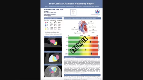
The newly cleared offering, AutoChamber, was designed with opportunistic screening in mind. It can evaluate many different kinds of CT images, including those originally gathered to screen patients for lung cancer. 

Trimed Popup
Trimed Popup