Cardiac Imaging

While cardiac ultrasound is the widely used imaging modality for heart assessments, computed tomography (CT), magnetic resonance imaging (MRI) and nuclear imaging are also used and are often complimentary, each offering specific details about the heart other modalities cannot. For this reason the clinical question being asked often determines the imaging test that will be used.

(Not so) average heart model created using CT images

Researchers at Pompeu Fabra University in Barcelona, Spain, have created a high-resolution, 3D atlas of the heart, providing a statistically average description of how the heart and its components look.

Small abdominal aortic aneurysms need less frequent ultrasound surveillance

In abdominal aortic aneurysm (AAA) screening programs, surveillance intervals of several years may be clinically acceptable for the majority of patients with small AAA since the smallest aneurysms often do not appear to change for many years, according to a meta-analysis published in the February 27 issue of the Journal of the American Medical Association.

Strong age relationship exists with coronary plaque burden, composition

The absence of coronary artery calcium (CAC) at CT strongly excludes patients from obstructive disease, while coronary atherosclerotic plaque burden is correlated with CAC scores in all ages, according to a study published in the February issue of Academic Radiology.

Thumbnail

Customized graft repair saves aneurysm patients from open surgery

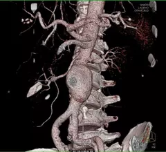
A newly approved graft, which is customized for individual patients suffering from abdominal aortic aneurysm using CT models, can provide an alternative to open surgery for some patients.

CCTA predicts heart attack risk for patients with no treatable risk factors

Coronary CT angiography (CCTA) is effective for determining the risk of heart attacks and other major adverse cardiovascular events in patients with suspected coronary artery disease (CAD) but no treatable risk factors, according to a study published Feb. 19 in Radiology.

Dual-source CT stacks up to 64-slice CT for hard-to-image patients

Newer dual-source cardiac CT scanners can provide accurate anatomic information to aid in the diagnosis and assessment of coronary artery disease (CAD) in patients who are difficult to image using older types of CT scanners, according to a study published online Feb. 7 in Radiology.

N.Y. hospital picks Merge's cardiology suite

Arnot Ogden Medical Center, a member of Arnot Health, will implement Merge Healthcare's cardiology suite to capture, manage and display multimodality cardiac images, as well as hemodynamics and ECG data across its enterprise.

FDA approves manufacturing site for Definity

The FDA has approved Jubilant HollisterStier as a manufacturing site for Lantheus Medical Imaging’s ultrasound imaging agent Definity Vial for Injectable Suspension.

Around the web

The nuclear imaging isotope shortage of molybdenum-99 may be over now that the sidelined reactor is restarting. ASNC's president says PET and new SPECT technologies helped cardiac imaging labs better weather the storm.

CMS has more than doubled the CCTA payment rate from $175 to $357.13. The move, expected to have a significant impact on the utilization of cardiac CT, received immediate praise from imaging specialists.

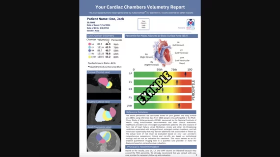
The newly cleared offering, AutoChamber, was designed with opportunistic screening in mind. It can evaluate many different kinds of CT images, including those originally gathered to screen patients for lung cancer. 

Trimed Popup
Trimed Popup