Computed Tomography

Computed tomography (CT) is a fast and accurate imaging modality often used in emergency settings and trauma imaging. CT scans, with or without (or both) iodinated contrast are frequently used to image the brain, chest, abdomen and pelvis, but also have post-imaging reconstructive capabilities for detailed orthopedic imaging. It is now a standard imaging modality in emergency rooms to quickly assess patients. CT uses a series of X-ray images shot as the gantry rotates around the patient. Computer technology assembles these into into a dataset volume than can be slices on any access, or advanced visualization software can extract specific parts of the anatomy for study. Find more content specific to cardiac CT.
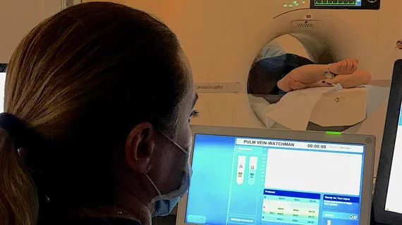
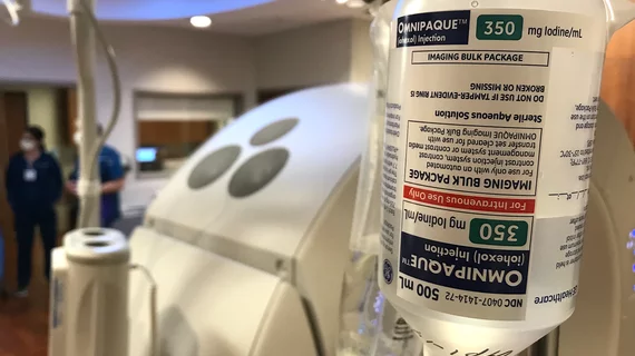
Deep learning recons reduce contrast and radiation doses in aortic CTA

Deep learning reconstruction cuts radiation and contrast dose by half in aortic CTA exams

A new analysis in in Academic Radiology dives into the importance of finding balance between reducing radiation exposure while maintaining image quality. 

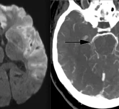
Dual energy CT parameters help determine the type of clot behind strokes

Knowledge of clot composition helps guide clinical management decisions following acute ischemic stroke.

Thumbnail

Is CT overused on patients involved in car crashes?

These exams can place an added burden on both patients and healthcare staffers, according to new research. 

Field Museum using CT scans to study mummified subjects.

Mobile CT unit helps Field Museum experts gain new insight into ancient history

Using computed tomography allows researchers to study these individuals in a more sensitive, person-centered approach.

Bayer's new CT contrast injection system earns FDA clearance.

New contrast injection system inks FDA clearance

The system can be set up in as little as two minutes and needs just 20 seconds to prime in between patients. 

‘A huge win’: CMS significantly increases Medicare payments for cardiac CT

CMS has more than doubled the CCTA payment rate from $175 to $357.13. The move, expected to have a significant impact on the utilization of cardiac CT, received immediate praise from imaging specialists.

HeartLung Technologies, a Houston-based artificial intelligence (AI) company, has received 510(k) clearance from the U.S. Food and Drug Administration (FDA) for new software that assesses chest CT scans for signs of coronary artery disease (CAD) and other potentially fatal heart conditions. The newly cleared offering, AutoChamber, was designed with opportunistic heart evaluations in mind.

FDA clears AI tool that flags signs of heart disease in chest CT scans

The newly cleared offering, AutoChamber, was designed with opportunistic screening in mind. It can evaluate many different kinds of CT images, including those originally gathered to screen patients for lung cancer. 

lung cancer screening

Patients without PCPs struggle to access lung cancer screening, despite eligibility

It is estimated that up to one-third of the U.S. population does not have a primary care provider.

Around the web

The nuclear imaging isotope shortage of molybdenum-99 may be over now that the sidelined reactor is restarting. ASNC's president says PET and new SPECT technologies helped cardiac imaging labs better weather the storm.

CMS has more than doubled the CCTA payment rate from $175 to $357.13. The move, expected to have a significant impact on the utilization of cardiac CT, received immediate praise from imaging specialists.

The newly cleared offering, AutoChamber, was designed with opportunistic screening in mind. It can evaluate many different kinds of CT images, including those originally gathered to screen patients for lung cancer. 

Trimed Popup
Trimed Popup