Computed Tomography

Computed tomography (CT) is a fast and accurate imaging modality often used in emergency settings and trauma imaging. CT scans, with or without (or both) iodinated contrast are frequently used to image the brain, chest, abdomen and pelvis, but also have post-imaging reconstructive capabilities for detailed orthopedic imaging. It is now a standard imaging modality in emergency rooms to quickly assess patients. CT uses a series of X-ray images shot as the gantry rotates around the patient. Computer technology assembles these into into a dataset volume than can be slices on any access, or advanced visualization software can extract specific parts of the anatomy for study. Find more content specific to cardiac CT.
artificial intelligence AI heart cardiology

Cleerly’s AI software for noninvasive FFR estimates receives a Category 1 CPT code

The company officially launched its Cleerly ISCHEMIA software for delivering noninvasive evaluations of CCTA exams in early 2024. This update provides a closer look at the software from a billing perspective. 

Examples of photon-counting coronary angiography showing how clarity improves as the thickness of the image is reduced. Top: 60-year-old female, with noncalcified plaque (arrowheads) and coronary stenosis (inset images). The reduced section thickness did not affect assessment in this patient. Bottom: 56-year-old female with calcified plaque (arrowheads) and coronary stenosis. The reduced section thickness leads to less calcium blooming and therefore a less severe percentage of stenosis. Courtesy of RSNA

Coronary artery disease classification improved with photon-counting CT

After a photon-counting CT, 54% of patients had their coronary artery disease classification downgraded.

Video of Mahadevappa Mahesh, PhD, incoming-AAPM president, professor of radiology and a medical physicist, Johns Hopkins University School of Medicine, explains key trends in imaging physics presented at the Radiological Society of North America (RSNA) 2023 meeting.

6 key trends in medical imaging physics

Mahadevappa Mahesh, PhD, incoming American Association of Physicists in Medicine president, discusses key developments in the specialty. 

Thumbnail

Micro-CT used to scan mysterious wormlike reptiles

The underground life of an Amphisbaenia has not been easy to study.

Thumbnail

Music eases anxiety during imaging. But, don’t let patients pick their playlist

Not all genres are equal in a clinical setting.

Thumbnail

Study examines outcomes of resection for ground-glass adenocarcinomas in the lungs

Researchers from Harvard Medical School looked into clinical findings for three types of nodules seen across 469 patients.

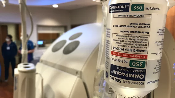
Supply crunch forces ED to develop contrast use mitigation protocol for PE

The protocol led to a reduction in CT scans without sacrificing diagnostic accuracy.

Kale Turtle Cook Museum of Natural Science

Rare sea turtle with infected shell undergoes CT scan

The procedure is medically necessary, but it also provides scientists insights into the Kemp’s ridley species of sea turtle.

Around the web

The nuclear imaging isotope shortage of molybdenum-99 may be over now that the sidelined reactor is restarting. ASNC's president says PET and new SPECT technologies helped cardiac imaging labs better weather the storm.

CMS has more than doubled the CCTA payment rate from $175 to $357.13. The move, expected to have a significant impact on the utilization of cardiac CT, received immediate praise from imaging specialists.

The newly cleared offering, AutoChamber, was designed with opportunistic screening in mind. It can evaluate many different kinds of CT images, including those originally gathered to screen patients for lung cancer. 

Trimed Popup
Trimed Popup