New software could improve diagnosis of lung conditions for patients who cannot tolerate contrast dye
New imaging software could provide more accurate diagnoses for patients with lung issues who are unable to receive contrast media during their CT scan.
Iodinated contrast media is a valuable tool in imaging, as it offers radiologists a more detailed view of patients’ anatomy. But for many people, contrast dye cannot be used due to either an allergy or medical condition that prohibits it. For those people, noncontrast exams are the only option, although they are known to be less accurate.
But new software developed by pulmonologist Girish Nair, MD, with Corewell Health William Beaumont University Hospital in Royal Oak, Michigan, and biomedical engineer Edward Castillo, PhD, with the University of Texas at Austin, could address the issue.
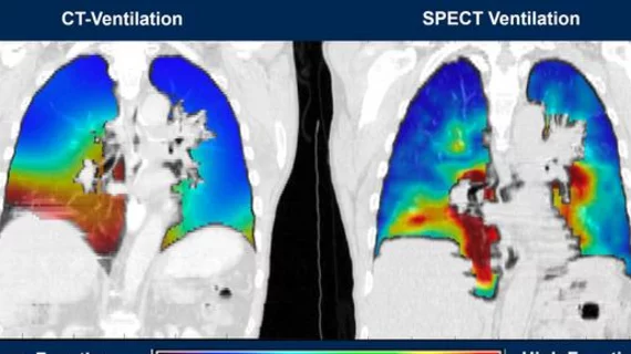
The machine learning-based software—CT-Derived Functional Imaging, or CTFI—uses a formula called the Integrated Jacobian Formulation to calculate lung volume based on metrics derived from imaging taken during inhalation and exhalation.
This sort of image capture is not new, though it is improved with this software, Nair indicated in a release.
“Up until now, a major problem with lung volume estimations from inhalation and exhalation CT scans were that results couldn’t be replicated for individual patients,” Nair said. “In other words, if you do a CT scan to calculate volume differences in the lungs the first time and then you try to do it again, you get different results every time for the same patient, making diagnosis and treatment harder.”
The software’s applicability extends beyond just those who cannot receive contrast. It can also improve the diagnostic process for patients suffering from chronic obstructive pulmonary disease and cancer, as it can help to target radiation therapy treatments. It can also limit the amount of radiation patients are exposed to.
“Directed radiation to treat a tumor is essential and if the patient is breathing in and out, then the tumor will naturally move or change position,” Nair explained. “This can result in the radiation going into other areas of the lung and potentially causing more harm than good.”
Data from research utilizing the software is set to be presented at the American Thoracic Society’s annual meeting this coming week in San Diego.

