Computed Tomography

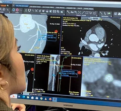
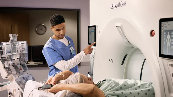
Computed tomography (CT) is a fast and accurate imaging modality often used in emergency settings and trauma imaging. CT scans, with or without (or both) iodinated contrast are frequently used to image the brain, chest, abdomen and pelvis, but also have post-imaging reconstructive capabilities for detailed orthopedic imaging. It is now a standard imaging modality in emergency rooms to quickly assess patients. CT uses a series of X-ray images shot as the gantry rotates around the patient. Computer technology assembles these into into a dataset volume than can be slices on any access, or advanced visualization software can extract specific parts of the anatomy for study. Find more content specific to cardiac CT.
Older adults among the walking wounded with incident TBI

The demographic most at risk seems to be White women who are healthy, active and of high socioeconomic status.

Thumbnail

Wake Forest receives $1.5 million to fund imaging study aimed at bone loss after bariatric surgery

The funding will go toward the Strategies to Reduce the Onset of Sleeve Gastrectomy Associated Bone Loss (STRONG BONES) trial.

Lung cancer screening CT image in a 66-year-old male patient shows a sessile nodule with internal air in the left mainstem to left upper lobe bronchus (arrow) with a mean diameter of 10 mm. The nodule was assigned as Lung-RADS category 4A in the clinical report. (B) Follow-up CT image shows the lesion is resolved. Image courtesy of RSNA

Lung-RADS update helps limit false-positive results, unnecessary procedures

Using the latest version was associated with improved diagnostic accuracy, researchers wrote in a new analysis. 

Dual energy CT scanners should be powered down more often to reduce energy consumption.

Why some CT equipment should be powered down more often

And not just on nights and weekends, authors of a new analysis on energy consumption in CT departments argue.

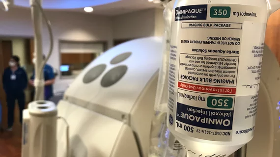
Investigation into patient's death after contrast injection begins

The man's wife told the court that he had undergone similar scans multiple times before and had never experienced any sort of adverse reaction. 

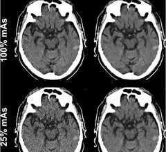
deep learning algorithm slashes radiation dose during neck CT scans

Deep learning denoising software can cut radiation exposure to 25% of conventional doses during neck CT

And the dose reduction can be achieved without sacrificing image quality, a new analysis shows.

AI software improves diagnosis of lung conditions when contrast cannot be used

New software could improve diagnosis of lung conditions for patients who cannot tolerate contrast dye

The machine learning-based software uses a formula called the Integrated Jacobian Formulation to calculate lung volume based on metrics derived from imaging taken during inhalation and exhalation. 

Thumbnail

Head CTs could present an opportunity to screen for osteoporosis

In individuals who have not completed a DEXA scan, head CT conducted for other reasons can offer insight into patients’ frontal bone density, a potential marker of osteoporosis.

Around the web

RBMA President Peter Moffatt discusses declining reimbursement rates, recruiting challenges and the role of artificial intelligence in transforming the industry.

Deepak Bhatt, MD, director of the Mount Sinai Fuster Heart Hospital and principal investigator of the TRANSFORM trial, explains an emerging technique for cardiac screening: combining coronary CT angiography with artificial intelligence for plaque analysis to create an approach similar to mammography.

A total of 16 cardiology practices from 12 states settled with the DOJ to resolve allegations they overbilled Medicare for imaging agents used to diagnose cardiovascular disease.