Computed Tomography

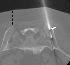
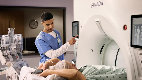
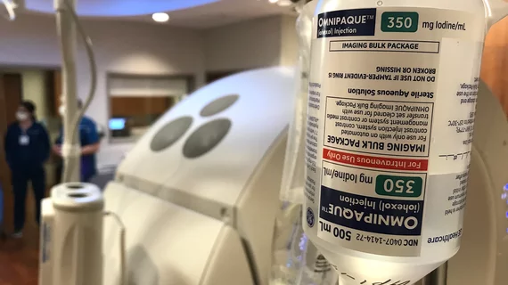
Computed tomography (CT) is a fast and accurate imaging modality often used in emergency settings and trauma imaging. CT scans, with or without (or both) iodinated contrast are frequently used to image the brain, chest, abdomen and pelvis, but also have post-imaging reconstructive capabilities for detailed orthopedic imaging. It is now a standard imaging modality in emergency rooms to quickly assess patients. CT uses a series of X-ray images shot as the gantry rotates around the patient. Computer technology assembles these into into a dataset volume than can be slices on any access, or advanced visualization software can extract specific parts of the anatomy for study. Find more content specific to cardiac CT.
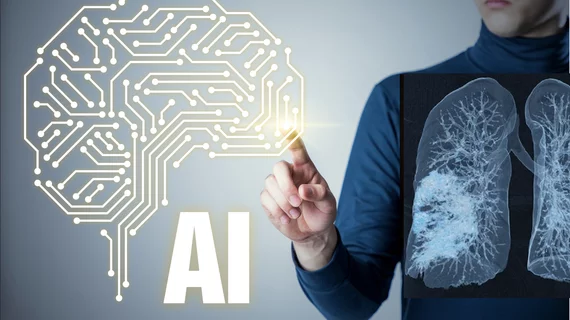
The rapid rise of artificial intelligence (AI) has helped cardiologists, radiologists, nurses and other healthcare providers embrace precision medicine in a way that ensures more heart patients are receiving personalized care.

FDA clears AI-enhanced 3D heart models generated from MR and CT images

The inHeart models significantly reduce cardiac ablation surgery times.

Head CT of a soldier with shrapnel wounds to head at a hospital in Dnipro. Photo by radiologist Olga Kachanova. Courtesy of RSNA.

PHOTO GALLERY: Radiology images of Ukrainian trauma patients wounded in war

A collection of medical images from hospitals in Ukraine showing radiology equipment in use and clinical images of wounded soldiers and civilians.


Micro-X mobile CT scanner

Mobile CT stroke detector passes VR simulation, set for field trials

The foldable unit weighs less than 154 pounds and can fit into an ambulance.

Radiologist using the Philips Smart Quant 3D Neuro artificial intelligence (AI) software to perform our measurements for white matter, gray matter and other parameters on brain MRI.

PHOTO GALLERY of brain imaging

This is a clinical photo gallery of neuro imaging and what conditions can be can be visualized in brain scans, and various imaging techniques used.

Outpatient study finds CT contrast is generally safe, but risk of adverse events varies with the seasons

A three-institution study of 473,482 patients found the risk of an adverse event following CT contrast increases in the summer and fall.

Medical 3D printing expert Summer Decker, PhD, professor and vice chair for research and innovation, and director of 3D clinical applications, University of South Florida (USF) Morsani College of Medicine and Tampa General Hospital, explains what is involved in creating a 3D printiung program in radiology and how it helps patient care.

Pitching the business case for 3D printing labs in radiology

Medical 3D printing expert Summer Decker, PhD, University of South Florida, explains how her lab has helped improve patient care. 

Thumbnail

New imaging protocols proposed to curb rise of cardiovascular infections

Eleven medical societies have signed on to a consensus statement aimed at standardizing imaging for suspected cardiovascular infections.

An MRI of a fetus inside the mother. The freakish appearance of the eyes and the face are normal for MRI fetal imaging. Image courtesy of RSNA

PHOTO GALLERY: What does fetal medical imaging look like?

This is a clinical photo gallery of fetal imaging that explains what all can be seen on medical imaging, how sex is determined, how measurements are used to track the development of a baby.

Around the web

The nuclear imaging isotope shortage of molybdenum-99 may be over now that the sidelined reactor is restarting. ASNC's president says PET and new SPECT technologies helped cardiac imaging labs better weather the storm.

CMS has more than doubled the CCTA payment rate from $175 to $357.13. The move, expected to have a significant impact on the utilization of cardiac CT, received immediate praise from imaging specialists.

The newly cleared offering, AutoChamber, was designed with opportunistic screening in mind. It can evaluate many different kinds of CT images, including those originally gathered to screen patients for lung cancer. 

Trimed Popup
Trimed Popup