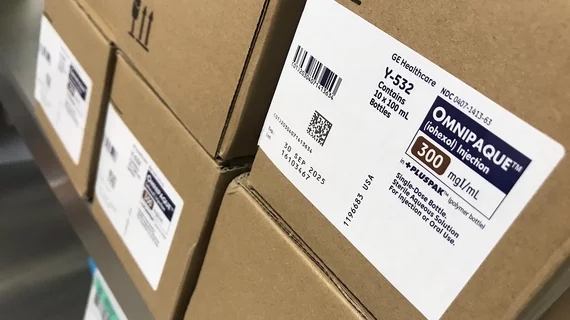
Imaging Contrast

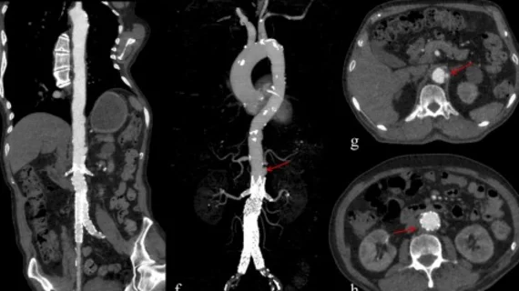
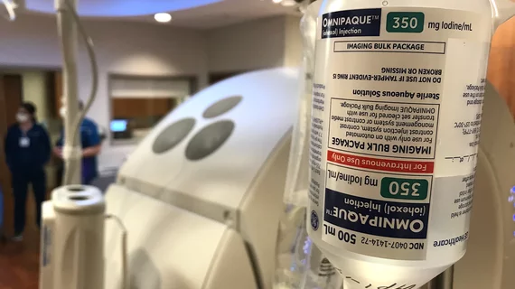
Contrast agents are injected into patients to help enhance images to make it easier for radiologists distinguish specific areas of the body from surrounding tissues. The most commonly used agents are iodinated contrast dye for computed tomography (CT), interventional cath lab angiography,  RF fluoroscopy, and in surgical OR procedures. MRI scans typically use gadolinium-based contrast agents. Ultrasound and echocardiography (cardiac ultrasound) imaging use contrast agents composed of microscopic bubbles to enhance images that otherwise would be suboptimal.

ASE recommends contrast agent usage in echoes

The American Society of Echocardiography (ASE) has issued a consensus statement, supporting the use of contrast agents used to enhance echocardiogram images, and providing a guide for physicians who may be hesitant to use the contrast agents following a 2007 FDA black-box warning. The statement was published in the November issue of Journal of the American Society of Echocardiography.

Around the web

The newly approved PET radiotracer is expected to improve patient care significantly. “We have been able to reach the pinnacle of myocardial perfusion imaging with flurpiridaz," one expert said.

GE HealthCare's flurpiridaz, the PET radiotracer that recently received FDA approval, offers several key benefits over SPECT. Jamshid Maddahi, MD, discussed the details in an exclusive interview. 

Ultrafast MCE could go on to become a go-to treatment option for obstructive coronary artery disease, according to the authors of a new first-in-human clinical study.

Trimed Popup
Trimed Popup