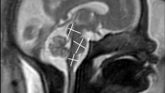
Fetal MRI safe on 3T scanners, new analysis shows
New research offers reassurance pertaining to the safety of fetal MRI exams conducted using 3T scanners.
MRI exams are crucial for identifying and investigating fetal abnormalities in utero, but in the past, concerns have been expressed about how the modality could affect intrauterine growth. Exams on 3T scanners account for around one-third of all fetal MRIs, and that number is expected to continue to increase.
With prior research on the topic being limited, it is important to get a better, more up-to-date understanding of whether the scan poses potential risk to a growing baby, authors of the new paper suggested.
“One concern is that the higher static magnetic field strength could influence biological processes, potentially leading to altered cellular functions or developmental anomalies,” corresponding author Teresa Victoria, MD, PhD, with the Department of Pediatric Imaging at Massachusetts General Hospital, and colleagues explained.
For the study, experts compared neonatal anthropometric measurements from newborns who underwent 3T fetal MRI, newborns who had 1.5T scans and newborns without any in utero MRI exposure. In total, there were 416 patients—104 in the 3T group, 104 in the 1.5T group and 208 in the control group.
On average, the MRIs were conducted between 25 and 27 weeks gestation. At birth, researchers did not observe significant differences in measurements, including weight, weight percentile, head circumference or head circumference percentile between the two groups.
The mean gestational age at delivery was lowest in the 3T MRI group, at 37 weeks 5 days. However, it did not vary substantially from the 1.5T group’s mean gestational age, at 38 weeks even, or the unexposed group’s, at 38 weeks 2 days.
“There has remained a lack of consensus in the radiology community on whether there is any actual risk of the use of 3T rather than 1.5T for fetal MRI,” the group noted, adding that that questions over safety have created a barrier to the widespread implementation of 3T fetal MRI exams, which provide superior diagnostic detail.
They suggested that their results further emphasize “the safety of 3-T MRI with respect to the growth of the developing fetus.”
The study abstract is available in the American Journal of Roentgenology.

