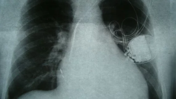
How MRI-safe are abandoned leads from cardiac implantable electronic devices?
As MRI technology continues to evolve, safety standards guiding use of the modality are generally lacking. This can make it difficult for patients and techs to determine whether an exam can be safely performed, especially when patients have older implantable devices.
Generally, things like cardiac implantable electronic devices (CIEDs) have long been considered a safety risk in MRI settings. But less is known about whether those risks remain when these items have been removed or replaced newer devices and their leads and/or lead fragments are left behind. Many newer devices are MRI conditional, but most legacy devices are not, leaving the question of whether their abandoned leads are hazardous when in the presence of a strong magnetic field.
“Patients with abandoned leads or lead fragments are considered at risk because of the unpredictable effects of lead tip heating during MRI and, as a secondary consideration, the risk associated with the CIED,” corresponding author Michael F. Morris, MD, with the department of radiology and division of cardiology at Banner University Medical Center Phoenix, and colleagues explain. “Although in vitro studies suggest that the length and orientation of fractured or abandoned leads contribute to heating during MRI, the effect in a clinical setting has not been reported. Additionally, it is unknown whether the length and orientation of abandoned leads or lead fragments affect CIED function in patients undergoing MRI.”
To better understand whether abandoned leads present additional risks, experts reviewed the medical charts of patients (126 total) with CIEDs and abandoned leads or lead fragments undergoing 1.5-T MRI from March 2014 through July 2020. Adverse clinical events during and up to 30 days after were analyzed, along with changes in CIED settings, such as composite threshold capture, sensing and lead impedance.
There were no reported deaths, clinically significant arrhythmias or other adverse clinical events in the group within 30 days of their scan. However, the CIEDs’ settings were affected during a small number of the exams. Three patients with abandoned leads had a significant change in composite of capture threshold, sensing, or lead impedance. They required CIED reprogramming, but no related adverse events were reported.
Further analysis did not reveal lead orientation, length, MRI type or scan duration to be associated with a significant change in the composite outcome, suggesting that adverse events in this population are rare when their exams are performed with 1.5T MRI scanners.
“In many instances, alternatives to MRI provide suboptimal diagnostic information. Alternatively, removing leads solely to facilitate MRI exposes patients to risks of morbidity and mortality,” the group explains, adding that their analysis supports consideration of 1.5-T MRI in this “historically contraindicated population.”
The study abstract can be found in Radiology: Cardiothoracic Imaging.

