
How patients' focus affects data derived from functional MRI scans
Functional MRI is a great way to observe the connectivity between different regions of the brain, offering experts insight into the processes that drive disease and human behavior. But new research suggests that waning focus during fMRI exams could be distorting the results.
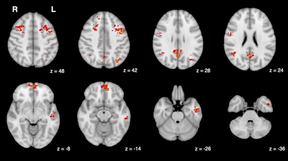
A group of experts from McLean Hospital, Harvard Medical School and the National Institute on Drug Abuse–Intramural Research Program (NIDA-IRP) discovered that the resultant brain activity observed on imaging could be misleading and potentially affect diagnoses.
“You’re laying down in a snug scanner for quite some time, often with only a low-engagement button press task to attend to or nothing to do at all, as the scanner monotonously hums and vibrates around you,” first author Cole Korponay, PhD, a research fellow at the McLean Hospital Imaging Center, notes. “These arousal-dampening conditions create the illusion that people’s brain connection strengths continuously inflate throughout the scan" to help better connect the ideas.”
Functional MR imaging uses changes in brain blood oxygen to measure neural activity. When patients become more relaxed or tired, their breathing and heart rates change. In turn, so does their blood oxygen. These changes can cause noise on imaging, making it difficult to get an accurate picture of brain activity.
The experts sought to identify a blood flow signal that could distinguish changes that are a result of decreased focus and legitimate fluctuations in functional connectivity. They discovered a non-neuronal, physiological noise signal that increased throughout patients’ scans in patterns that were in line with those observed during spikes in brain connectivity.
The team developed a denoising technique that effectively removes the “systemic low frequency oscillation” (sLFO) signal from fMRI data. This provides a more accurate depiction of connectivity strength, even during times when patients’ attention dwindles. This could improve the treatment of numerous conditions that rely on insight into brain connectivity patterns to determine patients’ treatment options, the group suggests.
“By adopting this sLFO denoising procedure, future studies can mitigate the distortive effects of arousal changes during brain scans and enhance the validity and reliability of fMRI findings,” Korponay indicates.
The study abstract is available in Nature Human Behavior.

In addition to her background in journalism, Hannah also has patient-facing experience in clinical settings, having spent more than 12 years working as a registered rad tech. She began covering the medical imaging industry for Innovate Healthcare in 2021.