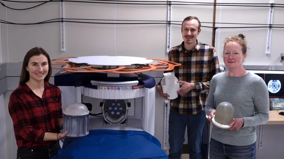
Researchers at the National Institute of Standards and Technology (NIST) are working on developing measurements that will provide better insight into soft tissue properties visualized on low-field MRI images.
Low-field MRI technology is an area many researchers have great interest in due to its ability to increase accessibility to the modality (many low-field units are portable) while also having the potential to decrease related costs. However, there is limited data on what tissue that is normal versus abnormal on low-field MRI looks like. Before these units can be widely deployed, experts need a better understanding of how to interpret low-field images.
“Magnetic resonance images of tissue differ depending on magnetic strength,” explained NIST electrical engineer Kalina Jordanova. “With low-field MRI systems, the contrast of the images is different, so we need to know how human tissue looks at these lower field strengths.”
Jordanova and colleagues recently sought to quantify tissue properties on low-field MRI exams as a means of creating a foundation for how these exams should be interpreted. To do this, they conducted brain scans on five male and five female volunteers using a commercially available portable MRI system with a magnetic field strength of 64 millitesla, which is significantly lower than that of conventional MR scanners.
The team imaged the entire brain, focusing on different areas that each produce distinct signals on imaging. This enabled them to gain a more quantitative understanding of tissue properties for gray matter, white matter and the patients' cerebrospinal fluid.
“Knowing the quantitative properties of tissue allows us to develop new image collection strategies for this MRI system,” NIST biomedical engineer Katy Keenan said.
The group is also studying how contrast agents affect low-field imaging, in addition to experimenting with the use of different materials that could potentially improve the image quality of low-field imaging.
To learn more, click here.

