
New sensor detects errors in MRI exams at 'lightning fast' speed
A new sensor capable of detecting subtle changes within a magnetic field could change the way MRI equipment is serviced in the future.
MRI equipment requires frequent calibration due to its fluctuating magnetic field, which can sometimes cause errors in imaging exams. But Hans Stærkind, a researcher at the Niels Bohr Institute and The Danish Research Center for Magnetic Resonance (DRCMR), and his team have developed a small sensor that can catch and correct these changes more quickly and potentially at a lower cost.
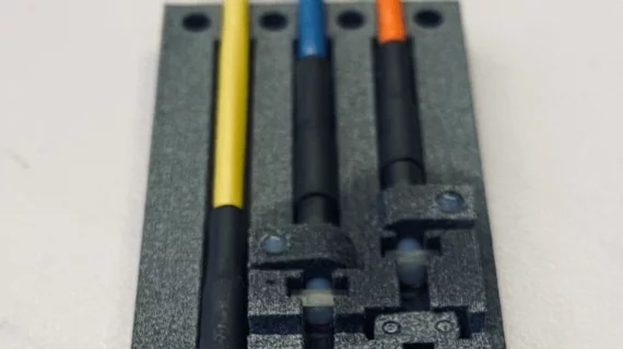
The sensor uses laser light encased in fiber cables and a small glass container filled with gas to measure changes in the strength of a magnetic field. It works by sending laser light through the fiber optic cables into four sensors placed within a scanner. Inside those sensors are small glass containers housing a cesium gas that absorbs light during certain frequencies.
"When the laser has just the right frequency while passing through the gas, there is a resonance between the waves of light and electrons in the cesium atoms,” Stærkind explained in a release. “But the frequency—or wavelength—at which this happens changes when the gas is exposed to a magnetic field. In this way, we can measure the strength of the magnetic field by finding out what the right frequency is."
Stærkind suggested that this happens “lighting fast” and can map out exactly where the disturbance in the magnetic field is occurring. In turn, this could enable users to quickly identify any magnetic field-related errors with exams in real-time, allowing them to make adjustments that could salvage images that may have previously been unusable.
The idea, Stærkind said, came from his late supervisor at DRCMR, Esben Petersen.
“He saw huge potential in developing a sensor based on lasers and gas that would be able to measure the magnetic fields without disturbing them," he said.
Staerkind developed the concept into a physical prototype—one that was made with hospitals in mind.
"The prototype is designed in such a way that it is already suitable in hospital contexts as a robust and well-functioning instrument. And so far, our tests have shown that it works as it should. One can imagine that this invention will eventually be integrated directly into new MRI scanners," he said.
Staerkind and his team are still working to fine-tune the sensor but are optimistic that one day it will be used to improve patient care.
"Once the prototype has been refined in a 2.0 version and its qualities documented with plenty of data from actual scans here at the hospital, we will see where this goes. It certainly has the potential to improve MRI scans in a unique way that can benefit doctors and, not least, patients.”

In addition to her background in journalism, Hannah also has patient-facing experience in clinical settings, having spent more than 12 years working as a registered rad tech. She began covering the medical imaging industry for Innovate Healthcare in 2021.