MRI finding linked to heightened cancer risk among women with very dense breasts
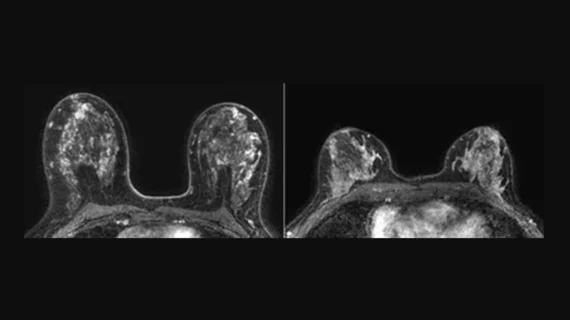
Higher volumes of background parenchyma enhancement (BPE) on MRI results are associated with a heightened cancer risk among women with very dense breasts, according to a new analysis published in Radiology.[1]
“Thus far, studies on breast cancer risk factors have typically focused on women at high lifetime risk of developing breast cancer,” co-author Kenneth G. A. Gilhuijs, PhD, a radiologist with the University Medical Center Utrecht in the Netherlands, said in a prepared statement from the Radiological Society of North America. “This is the first study that we know of that demonstrates an association between background parenchymal enhancement and occurrence of breast cancer in women with extremely dense breasts.”
Gilhuijs et al. developed an advanced artificial intelligence algorithm capable of identifying fibroglandular tissue using dynamic contrast-enhanced breast MRI exams from more than 4,500 patients. Exams were performed every two years from December 2011 to January 2016, and all data came from the Dense Tissue and Early Breast Neoplasm Screening (DENSE) trial.
A total of 122 patients were diagnosed with breast cancer over the course of the study. The researchers determined that breast cancer was twice as common among patients with the highest volumes of BPE than it was among women with the lowest volumes of BPE, highlighting the impact this finding could have on breast cancer screening strategies going forward.
“Our study is a first step in a direction to further tailor the frequency of supplemental MRI screening to individual women with dense breasts, focusing not only on breast density as a main risk factor but also on other properties of the breast established from a first screening MRI,” Gilhuijs said in the same statement.
The authors noted that they did not track race or ethnicity data for each patient. A future study that did include those details could help ensure any relationships between BPE and breast cancer risk are attributable to all demographics.
Click here to read the full Radiology study.

