Evidence points to optimal MRI sequence for detecting insufficiency fractures
New research has identified MRI sequences that could increase detection rates of insufficiency fractures that occur as a side effect of radiation therapy, which can be a diagnostic challenge to identify.
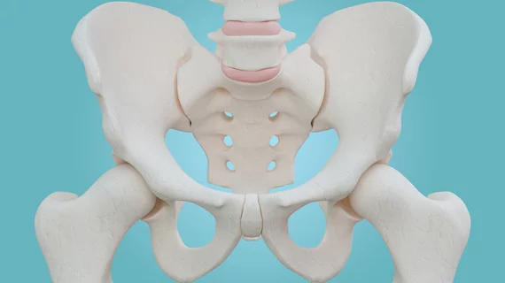
Sacral insufficiency fractures (SIFs) are a common side effect that women with cervical cancer are susceptible to after undergoing radiotherapy. But because SIFs do not always show a fracture line, they can be difficult to diagnose, or even misdiagnosed as bone metastasis, which could result in additional treatments like radio-chemotherapy. Therefore, it is important to optimize imaging sequences that can increase radiologist sensitivity to the signs of SIF, authors of the study noted.
“MRI is extraordinarily sensitive for revealing the reactive bone marrow edema associated with post-radiation IF and is useful for detecting soft-tissue mass, which may be valuable for identifying IF from bone metastasis, but MRI findings of insufficiency fractures may not always show a fracture line for SIF,” corresponding author Jiansheng Li, of the Department of Medical Imaging at Affiliated Cancer Hospital & Institute of Guangzhou Medical University in China, and co-authors discussed. “With the disappearance of fracture lines, the diagnosis of SIF is challenging.”
The authors explain that coronal fat saturation-T2W (FS-T2W) imaging of the sacrum increases detection rates of osteoporotic fractures and sacroiliitis by 6.8%, but that its utility in identifying radiation-induced SIFs had not been explored before their study was conducted. For their work, they compared coronal FS-T2W imaging with other MRI sequences to detail which best characterized SIF in cervical cancer patients who had undergone radiation therapy.
A cohort of 167 cervical cancer patients was narrowed down to 28 for whom SIFs were identified. MRI sequences compared were axial T1-weighted spin-echo images, axial T2-weighted spin-echo images and coronal fat-saturated T2-weighted images.
Fracture lines were detected in 64.6% of patients and lesion patterns were identified by assessing bone marrow edema patterns in all 28 individuals. For both, the experts found that the detection and characterization of coronal FS-T2WI outperformed T1WI and enhanced T1WI.
“We found coronal FS-T2WI was more sensitive to detect both bone marrow edema and fracture lines than that of T1WI or enhanced T1WI. In addition, this study revealed that T1WI detected the least fracture lines because the bone marrow edema pattern was also hypointensity, and enhanced T1WI detected least bone marrow edema due to some SIFs with no or mild enhancement, which may be difficult to identify,” the authors wrote, before concluding that this sequence could help avoid unnecessary and invasive treatment due to misdiagnosis.
The detailed research can be viewed in BMC Women’s Health.
Related orthopedic imaging content:
Researchers cite safety concerns after uncovering 'harmful behavior' of fracture-detecting AI model
AI assists radiologists in detecting fractures, improves workflow
Multi-slice knee MRI technique saves time without sacrificing quality
New guidance for knee cartilage MRI seeks to prevent irreversible osteoarthritis

