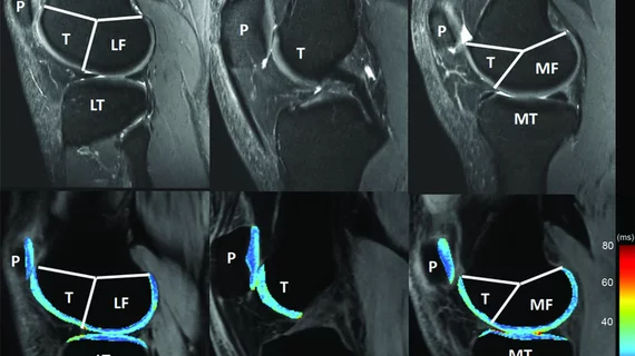
Performing serial MRIs in young patients with hemophilia may spot early joint damage
In young boys with hemophilia A, prompt and progressive treatment is necessary to delay the progression of joint deterioration. And new research suggests serial MRIs may play a vital role in detecting such damage.
Primary prophylaxis is considered the standard of care in boys with severe cases of the clotting disorder, but the effectiveness of treatment must be monitored. And MRI is considered the best tool for evaluating early soft tissue changes and osteochondral abnormalities.
For the study, researchers followed 56 boys managed across 11 hemophilia A treatment centers in Canada for more than 10 years. Participants consisted of boys with a starting age of 1- to 2-and-a-half-years-old. Researchers tracked interval and end-of-study changes noted on patients’ MRI scans.
During examinations, the boys underwent weekly prophylaxis infusions for a maximum of up to three times per week. MRIs were done on the boys’ ankles, elbows, and knees at the ages of 6- and 12-years-old to gauge the effectiveness of the infusions. Self-reported bleeding was also tracked during this time.
Two experienced radiologists, who were blinded to the clinical data, reviewed the images and noted that 54% of the boys showed detectable soft tissue damage in at least one joint. Synovial hypertrophy and hemosiderin deposition were also reported in 54% of participants. In conjunction with patients who self-reported bleeding, researchers established a 50% increase in the likelihood of joint deterioration.
“Soft-tissue changes in interval MRIs were associated with an increased risk of osteochondral findings in end-of-study MRIs and X-rays, suggesting that serial MRIs in populations of boys with hemophilia could be beneficial in guiding choice of individualized prophylaxis regimens aimed at minimizing subclinical and clinically overt joint bleeding and thus preserving long-term joint health,” Jennifer Stimec, MD, with the Department of Medical Imaging at The Hospital for Sick Children at the University of Toronto, and co-authors wrote.
While experts are still examining the overall efficacy of standard prophylaxis treatments, this study establishes the value of incorporating "serial MRIs” to detect early joint deterioration.
You can read the detailed results of the study in Research and Practice in Thrombosis and Haemostasis.

