Magnetic Resonance Imaging

Magnetic resonance imaging (MRI) is the gold standard imaging modality for soft tissues. It produces detail cross-sectional images of soft tissue and bone anatomy, including muscles, tendons, ligaments, brain and organs, without the use of ionizing radiation. In addition to orthopedic imaging, MRI is also used for heart, brain and breast. MRI uses gadolinium contrast in many exams to highlight tissues and blood vessels, which enhances images and offers better diagnostic quality. It can also be used in conjunction with PET scans. How does MRI work? MR creates images by using powerful magnets to polarize hydrogen atoms in water (the body is made of of more than 80% water) so they face in one direction. A radiofrequency pulse is then used to ping these atoms, causing them to wobble, or resonate. The MRI coils detect this and computers can assemble images from the signals. Basic MRI scans will focus on the resonance of fat and water in two different sequences, which highlight and contrast different features in the anatomy.

Thumbnail

Delays are common with sedated MRIs. Standardized workflow protocols could help avoid them

In some cases, MRI with sedation can delay workflows by 60 minutes or more, putting staff and patients in a bind.

How patients' focus affects data derived from functional MRI scans

As a person’s concentration dwindles during resting state or task-based sequences, the resultant brain activity observed on imaging could be misleading.

kid child pediatric MRI imaging

A lack of CPT codes for rapid pediatric MRI exams is costing patients

Abbreviated MRI exams for suspected appendicitis require fewer sequences and, in turn, take less time for radiologists to interpret. But in the absence of a designated code, patients often foot the bill for reads of more costly exams.

MRI could be key to diagnosing and treating depression

Researchers involved in the work are hopeful their findings will help patients find relief sooner.

Thumbnail

GBCA doses can be reduced by over 50% for some MRI exams

In some cases, contrast doses can be reduced by as much as 62% without sacrificing diagnostic quality.

Thumbnail

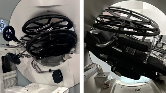
How MRI-safe are abandoned leads from cardiac implantable electronic devices?

Many newer devices are MRI conditional, but most legacy devices are not, leaving uncertainty around whether their abandoned leads are hazardous when in the presence of a strong magnetic field.

Thumbnail

Imaging research suggests Viagra could slow or prevent vascular dementia

A new study suggests sildenafil, known by its brand name Viagra, can improve cerebrovascular function and minimize the risk of cognitive impairment caused by small vessel disease. 

AI assistance improve prostate cancer margin definition

AI assistance yields prostate cancer margins 45x more accurate than those defined by rads alone

Even among the most seasoned urologists and radiologists, artificial intelligence software significantly improves prostate cancer mapping.

Around the web

The nuclear imaging isotope shortage of molybdenum-99 may be over now that the sidelined reactor is restarting. ASNC's president says PET and new SPECT technologies helped cardiac imaging labs better weather the storm.

CMS has more than doubled the CCTA payment rate from $175 to $357.13. The move, expected to have a significant impact on the utilization of cardiac CT, received immediate praise from imaging specialists.

The newly cleared offering, AutoChamber, was designed with opportunistic screening in mind. It can evaluate many different kinds of CT images, including those originally gathered to screen patients for lung cancer. 

Trimed Popup
Trimed Popup