Magnetic Resonance Imaging

Magnetic resonance imaging (MRI) is the gold standard imaging modality for soft tissues. It produces detail cross-sectional images of soft tissue and bone anatomy, including muscles, tendons, ligaments, brain and organs, without the use of ionizing radiation. In addition to orthopedic imaging, MRI is also used for heart, brain and breast. MRI uses gadolinium contrast in many exams to highlight tissues and blood vessels, which enhances images and offers better diagnostic quality. It can also be used in conjunction with PET scans. How does MRI work? MR creates images by using powerful magnets to polarize hydrogen atoms in water (the body is made of of more than 80% water) so they face in one direction. A radiofrequency pulse is then used to ping these atoms, causing them to wobble, or resonate. The MRI coils detect this and computers can assemble images from the signals. Basic MRI scans will focus on the resonance of fat and water in two different sequences, which highlight and contrast different features in the anatomy.

osseous lesion on MRI.

MRI scoring system could prevent unnecessary biopsy of osseous lesions

Radiation-induced osteitis may not cause clinical symptoms, but its presence often sparks concerns about malignancy.

Facial fillers seen on MRI

Imaging of woman with facial fillers prompts questions about whether they actually dissolve over time

Cosmetic facial fillers made from hyaluronic acid might not dissolve as quickly as previously believed, warns a California plastic surgeon. 

Thumbnail

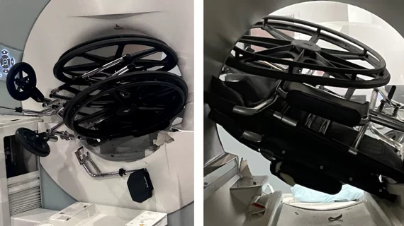
'A very, very good thing'—CMS proposes reimbursements for providers' efforts to ensure MRI safety

“Before these CPT codes there was no real acknowledgment of the additional burden borne by the providers who accepted these patients."

Multiple sclerosis lesions detected with AI assistance

AI assistance helps rads shave 1/3 of their reporting times for MS lesions

Assessing multiple sclerosis is a time-consuming process, making reducing the burden an interest of multiple AI vendors.

Thumbnail

Lululemon has no place in an MRI suite, doc warns

A neurosurgeon is cautioning patients to check the material makeup of their clothes before donning them during an MRI exam.

MRI study shows how magic mushrooms affect brain connectivity in a way that allows people to temporarily escape reality

MRIs pinpoint how magic mushrooms affect brain connectivity, paving way for effective mental health therapeutics

A new MRI analysis shows how psilocybin temporarily alters the signals of the brain’s default mode network.

Thumbnail

Structured reporting improves outcomes for patients with placenta accreta spectrum

New data details an association between the implementation of structured templates and a reduction in patients being admitted to the intensive care unit. 

Thumbnail

Men at risk for prostate cancer could be forgoing necessary imaging due to climbing out-of-pocket costs

Experts are calling for insurance companies to provide full coverage for imaging and other necessary exams, if indicated by an abnormal prostate cancer screening test.

Around the web

The newly approved PET radiotracer is expected to improve patient care significantly. “We have been able to reach the pinnacle of myocardial perfusion imaging with flurpiridaz," one expert said.

GE HealthCare's flurpiridaz, the PET radiotracer that recently received FDA approval, offers several key benefits over SPECT. Jamshid Maddahi, MD, discussed the details in an exclusive interview. 

Ultrafast MCE could go on to become a go-to treatment option for obstructive coronary artery disease, according to the authors of a new first-in-human clinical study.

Trimed Popup
Trimed Popup