Magnetic Resonance Imaging

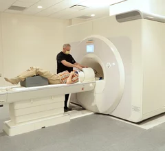
Magnetic resonance imaging (MRI) is the gold standard imaging modality for soft tissues. It produces detail cross-sectional images of soft tissue and bone anatomy, including muscles, tendons, ligaments, brain and organs, without the use of ionizing radiation. In addition to orthopedic imaging, MRI is also used for heart, brain and breast. MRI uses gadolinium contrast in many exams to highlight tissues and blood vessels, which enhances images and offers better diagnostic quality. It can also be used in conjunction with PET scans. How does MRI work? MR creates images by using powerful magnets to polarize hydrogen atoms in water (the body is made of of more than 80% water) so they face in one direction. A radiofrequency pulse is then used to ping these atoms, causing them to wobble, or resonate. The MRI coils detect this and computers can assemble images from the signals. Basic MRI scans will focus on the resonance of fat and water in two different sequences, which highlight and contrast different features in the anatomy.

Thumbnail

Bilingual people have more efficient brains, imaging study shows

Researchers examined the brains of bilingual and monolingual people using fMRI, only to find improved communication between isolated regions in those who learn a second language at a young age.

long covid brainstem damage

Ultra-high field MRI implicates long COVID in brainstem damage

Researchers have used 7T MRI to demonstrate that former COVID-19 inpatients are susceptible to persistent brainstem abnormalities associated with long-haul COVID symptoms.

Thumbnail

Outdated manuals pose MRI safety risks for patients with medical implants

After reviewing years of data from its clinic, one institution discovered that issues with implant data integrity frequently put patients at risk. 

Thumbnail

GPT-4 as accurate as neurologists in predicting final diagnosis based on MRI reports

The large language model can also outperform other human providers, radiologists included, new study shows.

Novel imaging technique could be used to tailor glioblastoma treatment

It allows providers to quantify changes in tumor size throughout patients’ treatment in real-time, providing early indications of efficacy and enabling adjustments as needed.

Thumbnail

MRI-guided biopsy decisions halve the rate of clinically insignificant prostate cancer diagnoses

“This paper is the message to healthcare authorities around the world to look over recommendations for men."

Portable MRI system slashes the time it takes to image stroke patients

Experts are optimistic that the use of portable systems can increase accessibility to neuroimaging and efficiency in stroke care. 

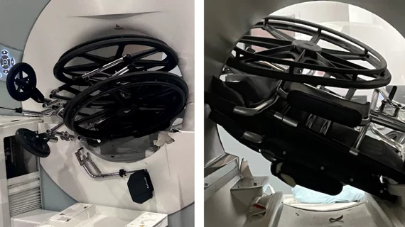
Imaging center drug raid goes awry after cop's gun is sucked into MRI scanner

A new lawsuit alleges that the LAPD staked out an imaging center due to its higher than usual energy consumption and a “distinct odor” of cannabis coming from the facility. 

Around the web

The newly approved PET radiotracer is expected to improve patient care significantly. “We have been able to reach the pinnacle of myocardial perfusion imaging with flurpiridaz," one expert said.

GE HealthCare's flurpiridaz, the PET radiotracer that recently received FDA approval, offers several key benefits over SPECT. Jamshid Maddahi, MD, discussed the details in an exclusive interview. 

Ultrafast MCE could go on to become a go-to treatment option for obstructive coronary artery disease, according to the authors of a new first-in-human clinical study.

Trimed Popup
Trimed Popup