Magnetic Resonance Imaging

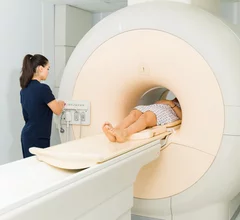
Magnetic resonance imaging (MRI) is the gold standard imaging modality for soft tissues. It produces detail cross-sectional images of soft tissue and bone anatomy, including muscles, tendons, ligaments, brain and organs, without the use of ionizing radiation. In addition to orthopedic imaging, MRI is also used for heart, brain and breast. MRI uses gadolinium contrast in many exams to highlight tissues and blood vessels, which enhances images and offers better diagnostic quality. It can also be used in conjunction with PET scans. How does MRI work? MR creates images by using powerful magnets to polarize hydrogen atoms in water (the body is made of of more than 80% water) so they face in one direction. A radiofrequency pulse is then used to ping these atoms, causing them to wobble, or resonate. The MRI coils detect this and computers can assemble images from the signals. Basic MRI scans will focus on the resonance of fat and water in two different sequences, which highlight and contrast different features in the anatomy.

Thumbnail

New MRI study contradicts prior research linking dementia with benzodiazepine use

There has long been speculation suggesting that chronic benzo consumption could be a precursor to conditions that deteriorate neurological function.

ping pong improve brain function

Playing ping pong improves brain connectivity

New research highlights key MRI findings related to connectivity and white matter integrity in the brains of athletes taking part in competitive table tennis.

Thumbnail

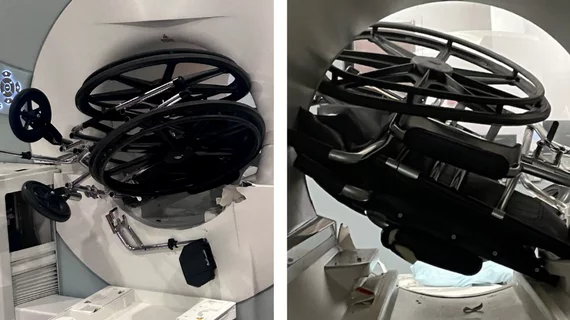
MRI safety concerns prompt FDA to issue recall for series of ventilators

The Class I recall affects the ZOLL 731 Series Ventilator family, including the AEV, EMV+, and Eagle II models.

Deep learning application spots acute ischemic stroke in less than 30 seconds on MRI

What’s more, the application’s performance was accurate, even when fewer scanning sequences were utilized.

Thumbnail

MRI findings predict Parkinson's disease progression

MR imaging could provide insight into how Parkinson’s disease will progress years before its related symptoms become debilitating.  

Murderer's fate hinges on 2 different interpretations of his brain scans

A radiologist’s testimony could deconstruct a legal team’s argument that their defendant had a brain tumor that may have affected his behavior when he shot and killed five women in 2019. 

Thumbnail

How having ovaries removed before menopause affects brain structure

These changes are especially prevalent in women who have the surgery before the age of 40, new research suggests. 

Thumbnail

Fluorinated contrast agents show 'strong potential' for MRI theranostics

Experts have developed a first-of-its-kind fluorinated contrast agent they believe will provide more detailed MR imaging and aid in the treatment of certain cancers. 

Around the web

The nuclear imaging isotope shortage of molybdenum-99 may be over now that the sidelined reactor is restarting. ASNC's president says PET and new SPECT technologies helped cardiac imaging labs better weather the storm.

CMS has more than doubled the CCTA payment rate from $175 to $357.13. The move, expected to have a significant impact on the utilization of cardiac CT, received immediate praise from imaging specialists.

The newly cleared offering, AutoChamber, was designed with opportunistic screening in mind. It can evaluate many different kinds of CT images, including those originally gathered to screen patients for lung cancer. 

Trimed Popup
Trimed Popup