
New imaging technique could change how Crohn's disease is treated
A new PET/MR imaging technique could pave the way for more targeted treatments for individuals suffering from Crohn's disease, according to a recent study.
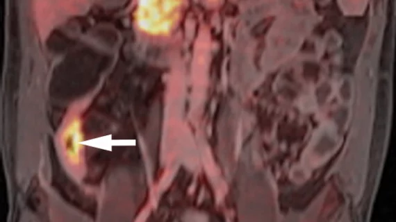
Published in Radiology, the new research details the use of a gallium 68 fibroblast activation protein inhibitor (FAPI) tracer for differentiating between inflammation of the intestinal wall and fibrosis. For patients with Crohn’s, distinguishing between the two is important because inflammatory strictures typically respond to noninvasive treatment, whereas fibrotic narrowing requires surgical intervention. Until recently, imaging exams were not able to offer the level of detailed information needed to determine whether patients’ symptoms were due to inflammation or fibrosis, authors of the new paper explained.
“Currently used cross-sectional imaging techniques enable reliable assessment of active transmural inflammation and identification of intestinal strictures in patients with Crohn's disease,” study co-leader Michael Bergmann, from MedUni Vienna's Department of General Surgery, and co-authors noted. “However, a correct estimation of tissue composition, including fibrosis, and the differentiation between inflammation and fibrosis remains a challenge for US and MRI.”
The FAPI radiotracer experts used in this research can bind specifically to the fibroblast activating protein of connective tissue cells that cause intestinal wall fibrosis, thus enabling clinicians to better differentiate fibrosis from active inflammation.
The team tested the radiotracer on 14 patients with Crohn’s disease and obstructive symptoms. Each patient underwent preoperative 68Ga-FAPI PET/MR enterography and postoperative histological analysis to grade inflammation and fibrosis; these results were then compared to patients’ PET/MRI findings.
Through this, researchers observed higher maximum standard uptake values (SUVmax) of the radiotracer in bowel segments with fibrosis in comparison to segments without it. Additionally, segments with isolated active inflammation showed lower uptake than segments where experts observed a combination of inflammation and fibrosis.
These findings could be used to guide patients and providers in making decisions relative to treatment strategies, the authors suggested.
"In [the] future, the molecular imaging we have developed could be used to identify those patients who would benefit from surgical intervention at an early stage, thereby sparing them the need for less effective drug therapy for fibroid-stenosis," Bergmann noted.
Follow-up studies using the radiotracer are already in the works. Those studies will be conducted on a larger scale and also will investigate the progression of fibroid-stenosis and the effects various therapies have on the disease.
The study abstract is available here.

In addition to her background in journalism, Hannah also has patient-facing experience in clinical settings, having spent more than 12 years working as a registered rad tech. She began covering the medical imaging industry for Innovate Healthcare in 2021.