Molecular Imaging

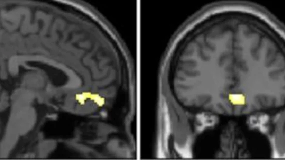
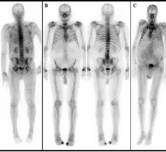
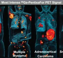
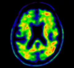
Molecular imaging (also called nuclear medicine or nuclear imaging) can image the function of cells inside the body at the molecular level. This includes the imaging modalities of positron emission computed tomography (PET) and single photon emission computed tomography (SPECT) imaging. How does PET and SPECT imaging work? Small amounts of radioactive material (radiopharmaceuticals) injected into a patient. These can use sugars or chemical traits to bond to specific cells. The radioactive material is taken up by cells that consume the sugars. The radiation emitted from inside the body is detected by photon detectors outside the body. Computers take the data to assemble images of the radiation emissions. Nuclear images may appear fuzzy or ghostly rather than the sharper resolution from MRI and CT.  But, it provides metabolic information at a cellular level, showing if there are defects in the function of the heart, areas of very high metabolic activity associated with cancer cells, or areas of inflammation, data not available from other modalities. These noninvasive imaging exams are used to diagnose cancer, heart disease, Alzheimer’s and Parkinson’s disease, bone disorders and other disorders. 

lung cancer pulmonary nodule chest

Preoperative PET/CT imaging linked with better survival in advanced lung cancer

PET/CT imaging in these patients increases overall survival depending on the cancer’s stage, with those diagnosed with stage 3A and 3B NSCLC appearing to benefit the most from the exam. 

U.S. News & World Report children’s hospitals cardiology heart surgery

Lower doses, faster acquisitions: Experts share how to improve PET scans for peds

This week in AJR, experts shared how they were able to reduce FDG PET scan acquisition times by 33%, thus reducing the amount of total radiation exposure to more vulnerable pediatric patients. 

Targeted radionuclide alpha therapy study wins SNMMI's Abstract of the Year

The study touts the benefits of targeted radionuclide alpha therapy for patients with gastroenteropancreatic neuroendocrine tumors (GEP-NETs) who have not had success with other treatments.

PSMA PET mapping improves radiation therapy contouring after PCA recurrence

Salvage radiation therapy guidelines for patients with prostate cancer recurrence need to be updated to include findings identified on PSMA PET, according to new research shared at SNMMI’s annual conference this week. 

Siemens debuts new, cleared SPECT/CT model

Siemens Healthineers splashed an FDA-approved SPECT/CT system June 12 at the 2022 annual meeting of the Society of Nuclear Medicine & Nuclear Imaging in Vancouver, B.C.

Thumbnail

Cutting radiation exposure in half during PET/CT using virtual CT scans

“High-quality artificial intelligence-generated images preserve vital information from raw PET images without the additional radiation exposure from CT scans,” experts involved in the study explained.

Thumbnail

Wisconsin company to begin producing medical isotopes next year

The company’s CEO, Greg Piefer, detailed the plans at the Wisconsin Entrepreneurs’ Conference on June 2, stating that production would be achieved via small-scale nuclear transmutation.

Thumbnail

One chemistry professor's role in increasing radioisotope production in the U.S.

The work of a chemistry professor at the University of Wisconsin-Milwaukee is helping to blaze the trail of reducing the United States’ dependence on foreign countries for vital medical isotopes. 

Around the web

GE HealthCare's flurpiridaz, the PET radiotracer that recently received FDA approval, offers several key benefits over SPECT. Jamshid Maddahi, MD, discussed the details in an exclusive interview. 

Ultrafast MCE could go on to become a go-to treatment option for obstructive coronary artery disease, according to the authors of a new first-in-human clinical study.

Elucid's PlaqueIQ was trained to turn CCTA images into interactive 3D reports that help physicians visualize the presence of atherosclerosis.

Trimed Popup
Trimed Popup