Molecular Imaging

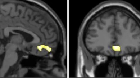
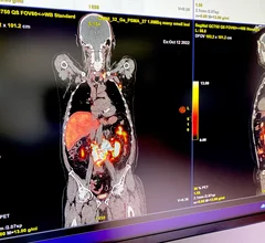
Molecular imaging (also called nuclear medicine or nuclear imaging) can image the function of cells inside the body at the molecular level. This includes the imaging modalities of positron emission computed tomography (PET) and single photon emission computed tomography (SPECT) imaging. How does PET and SPECT imaging work? Small amounts of radioactive material (radiopharmaceuticals) injected into a patient. These can use sugars or chemical traits to bond to specific cells. The radioactive material is taken up by cells that consume the sugars. The radiation emitted from inside the body is detected by photon detectors outside the body. Computers take the data to assemble images of the radiation emissions. Nuclear images may appear fuzzy or ghostly rather than the sharper resolution from MRI and CT.  But, it provides metabolic information at a cellular level, showing if there are defects in the function of the heart, areas of very high metabolic activity associated with cancer cells, or areas of inflammation, data not available from other modalities. These noninvasive imaging exams are used to diagnose cancer, heart disease, Alzheimer’s and Parkinson’s disease, bone disorders and other disorders. 

Thumbnail

'Quite impressive': ChatGPT generates a nuclear medicine report

The generated report included indication, findings laid out numerically, TNM stage, impression and follow-up recommendations.

New practice guidelines for PET imaging of neuroendocrine tumors

Issued by the Society of Nuclear Medicine and Molecular Imaging (SNMMI) and the European Association of Nuclear Medicine (EANM), the new guidelines are intended to inform any practitioners involved in ordering, performing, reading and reporting SSTR PET imaging.

Thumbnail

Amyloid plaque patterns on PET imaging predict Alzheimer's progression in asymptomatic patients

Experts involved in the new research suggest that identifying these spatiotemporal variations could play an important role in clinical research and precision medicine. 

Thumbnail

E-cigarettes cause worse inflammation than tobacco cigarettes, new PET data shows

The use of e-cigarettes has increased significantly in recent years, mostly due to the fact that many consumers believe them to be a safer option than traditional cigarettes.

Thumbnail

Advanced PET/MRI protocols boost lung cancer detection

Experts involved in the new research indicate that PET/MRI could have an increasingly prevalent role in lung cancer staging and restaging.

new radiotracer studies inflammation

New radiotracer could change how radiologists understand inflammation

Labeled as LW223, the fluorinated PET radiotracer is the first of its kind to be impervious to rs6971 polymorphism—a genetic mutation that inhibits the diagnostic view of inflammation imaging.

vaccine syringe covid-19 coronavirus

PET radiotracer reduces false positives owed to COVID vaccination

With COVID booster vaccinations expected to continue, the new findings are especially relevant.

Thumbnail

Plans for another nuclear facility that can produce medical isotopes in the works

Many of the facilities that produce these critical isotopes are aging, which is a big cause for concern in the future among experts in the field, many of whom believe that the supply chain for radioisotopes is already fragile.

Around the web

GE HealthCare's flurpiridaz, the PET radiotracer that recently received FDA approval, offers several key benefits over SPECT. Jamshid Maddahi, MD, discussed the details in an exclusive interview. 

Ultrafast MCE could go on to become a go-to treatment option for obstructive coronary artery disease, according to the authors of a new first-in-human clinical study.

Elucid's PlaqueIQ was trained to turn CCTA images into interactive 3D reports that help physicians visualize the presence of atherosclerosis.

Trimed Popup
Trimed Popup