Molecular Imaging

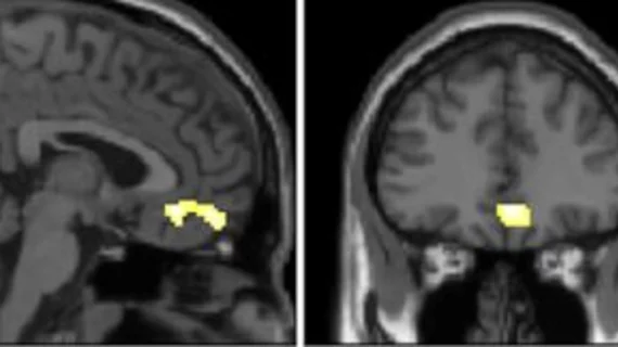
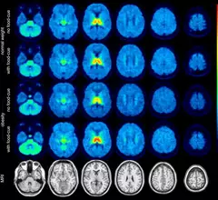
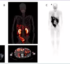
Molecular imaging (also called nuclear medicine or nuclear imaging) can image the function of cells inside the body at the molecular level. This includes the imaging modalities of positron emission computed tomography (PET) and single photon emission computed tomography (SPECT) imaging. How does PET and SPECT imaging work? Small amounts of radioactive material (radiopharmaceuticals) injected into a patient. These can use sugars or chemical traits to bond to specific cells. The radioactive material is taken up by cells that consume the sugars. The radiation emitted from inside the body is detected by photon detectors outside the body. Computers take the data to assemble images of the radiation emissions. Nuclear images may appear fuzzy or ghostly rather than the sharper resolution from MRI and CT.  But, it provides metabolic information at a cellular level, showing if there are defects in the function of the heart, areas of very high metabolic activity associated with cancer cells, or areas of inflammation, data not available from other modalities. These noninvasive imaging exams are used to diagnose cancer, heart disease, Alzheimer’s and Parkinson’s disease, bone disorders and other disorders. 

cardiac amyloidosis on bone scan

Cardiac amyloidosis becoming less rare thanks to nuclear medicine studies

A new study in the Journal of Nuclear Medicine offers insight into how the condition affects the general population, as well as how radiologists can help in the cardiac amyloidosis diagnostic journey.

Nuclear Regulatory Commission to reconsider extravasation reporting requirements

The NRC noted that advancements in nuclear medicine and increased use of radiopharmaceuticals prompted the commission to reconsider the exclusion of extravasation from medical event reporting.  

Thumbnail

CMS cites 'newly published evidence' in postponement of PET coverage decision

Prior to the Dec. 15 announcement, CMS had released a different statement in June informing the public that they were reconsidering the amount of PET scans that Alzheimer’s patients undergoing monoclonal antibody treatment can receive.

Thumbnail

U.S. lawmakers pen letter calling for expanded access to amyloid PET imaging

“We must make every effort to grant equitable access to tools that can help individuals and their loved ones treat and manage this disease."

New radiotracer IDs numerous cancers likely to respond to targeted therapy

The radiotracer, 68Ga-PentixaFor, can be used for detecting C-X-C motif chemokine receptor 4 (CXCR4), which plays a significant role in cancer progression.

Thumbnail

Cognitive decline not far behind amyloid and tau PET-positive scans, even for the asymptomatic

Common markers of Alzheimer's disease visualized on PET scans of asymptomatic patients might foreshadow the onset of symptoms in the years following their initial discovery.

Thumbnail

How a newly secured 3D printing patent could stabilize radioisotope production

Recently, a patent for 3D printed uranium targets was secured by the National Center for Nuclear Research (NCBJ) in Swierk, Poland, and it could increase the production of Molybdenum-99 (Mo-99) globally. 

NorthStar radiopharmaceutical development facility.

NorthStar breaks ground on new facility, will offer radiopharmaceutical development services to other companies

The 36,000 square foot unit will be located in Beloit, Wis., alongside NorthStar’s production facilities. It is expected to be open for business starting in 2024.

Around the web

GE HealthCare's flurpiridaz, the PET radiotracer that recently received FDA approval, offers several key benefits over SPECT. Jamshid Maddahi, MD, discussed the details in an exclusive interview. 

Ultrafast MCE could go on to become a go-to treatment option for obstructive coronary artery disease, according to the authors of a new first-in-human clinical study.

Elucid's PlaqueIQ was trained to turn CCTA images into interactive 3D reports that help physicians visualize the presence of atherosclerosis.

Trimed Popup
Trimed Popup