Molecular Imaging

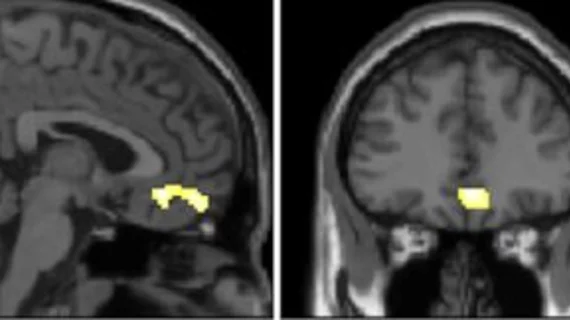
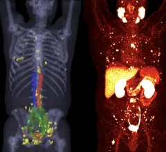
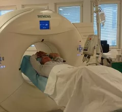
Molecular imaging (also called nuclear medicine or nuclear imaging) can image the function of cells inside the body at the molecular level. This includes the imaging modalities of positron emission computed tomography (PET) and single photon emission computed tomography (SPECT) imaging. How does PET and SPECT imaging work? Small amounts of radioactive material (radiopharmaceuticals) injected into a patient. These can use sugars or chemical traits to bond to specific cells. The radioactive material is taken up by cells that consume the sugars. The radiation emitted from inside the body is detected by photon detectors outside the body. Computers take the data to assemble images of the radiation emissions. Nuclear images may appear fuzzy or ghostly rather than the sharper resolution from MRI and CT.  But, it provides metabolic information at a cellular level, showing if there are defects in the function of the heart, areas of very high metabolic activity associated with cancer cells, or areas of inflammation, data not available from other modalities. These noninvasive imaging exams are used to diagnose cancer, heart disease, Alzheimer’s and Parkinson’s disease, bone disorders and other disorders. 

Thumbnail

Isotope shortage update: NMEU names potential restart date for downed reactor

On November 4, Nuclear Medicine Europe indicated inspections at the downed BR2 reactor had been finalized.

Thumbnail

Another isotope shortage looms as mechanical failure delays production

A new report suggests there may soon be a shortage of molybdenum-99 (Mo-99)/technetium-99m (Tc-99m). What does that mean for patient care in the months ahead? 

Barry L. Zaret, MD

Veteran cardiologist, viewed by many as the founder of nuclear cardiology, dies at 82

Barry L. Zaret, MD, was an influential cardiologist, accomplished poet and veteran of the United States Air Force. 

Thumbnail

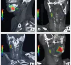
PET/CT findings predict post-treatment, radiation-induced hypothyroidism

Radiation-induced hypothyroidism is common yet underdiagnosed, potentially owing to a lack of follow-up consensus in patients treated with radiation therapy for head and neck cancers.

Thumbnail

FAPI PET/CT findings linked with risk of sudden cardiac death

Experts involved in the research suggested that these findings indicate a potential role for FAPI PET/CT imaging in detecting changes in myocardial fibrosis, noting that it could be more sensitive to earlier changes than standard cardiac MRI.

FAPI PET/CT shows promise as a better radiotracer for cancer than 18F-FDG. It also can be used for detection of infection and inflammation.

VIDEO: 6 key advances in molecular imaging technology for PET and SPECT

Munir Ghesani, MD, President of the Society of Nuclear Medicine and Molecular Imaging (SNMMI), system chief of nuclear medicine at Mount Sinai Health, explains recent advances in nuclear imaging technology.

Thumbnail

A new Alzheimer's drug therapy appears effective. What might this mean for the future of amyloid PET?

Previously, CMS determined that coverage for patients receiving treatment was dependent on their being enrolled in a CMS-approved clinical trial under coverage with evidence development (CED).

Thumbnail

Does the use of PSMA PET impact tumor upstaging?

A new risk analysis suggests the exam will increasingly result in tumor upstaging among high-risk patients.

Around the web

GE HealthCare's flurpiridaz, the PET radiotracer that recently received FDA approval, offers several key benefits over SPECT. Jamshid Maddahi, MD, discussed the details in an exclusive interview. 

Ultrafast MCE could go on to become a go-to treatment option for obstructive coronary artery disease, according to the authors of a new first-in-human clinical study.

Elucid's PlaqueIQ was trained to turn CCTA images into interactive 3D reports that help physicians visualize the presence of atherosclerosis.

Trimed Popup
Trimed Popup