Molecular Imaging

Molecular imaging (also called nuclear medicine or nuclear imaging) can image the function of cells inside the body at the molecular level. This includes the imaging modalities of positron emission computed tomography (PET) and single photon emission computed tomography (SPECT) imaging. How does PET and SPECT imaging work? Small amounts of radioactive material (radiopharmaceuticals) injected into a patient. These can use sugars or chemical traits to bond to specific cells. The radioactive material is taken up by cells that consume the sugars. The radiation emitted from inside the body is detected by photon detectors outside the body. Computers take the data to assemble images of the radiation emissions. Nuclear images may appear fuzzy or ghostly rather than the sharper resolution from MRI and CT.  But, it provides metabolic information at a cellular level, showing if there are defects in the function of the heart, areas of very high metabolic activity associated with cancer cells, or areas of inflammation, data not available from other modalities. These noninvasive imaging exams are used to diagnose cancer, heart disease, Alzheimer’s and Parkinson’s disease, bone disorders and other disorders. 

Thumbnail

SNMMI names 2014-2016 Wagner-Torizuka fellows

The Wagner-Torizuka fellowship, created by Henry N. Wagner, Jr., MD, and Kanji Torizuka, MD, PhD, provides Japanese clinicians in the field of molecular imaging a major platform for advanced training and research. Three physicians have been honored with the fellowship, SNMMI announced yesterday. 

Novel biomarker looks for diabetic retinopathy

A targeted molecular imaging technique could aid in the prevention of blindness associated with progressive diabetic neuropathy by tracking alterations in the retinal endothelium, the Federation of American Societies for Experimental Biology (FASEB) announced yesterday.

Thumbnail

Vizamyl wins European approval

The amyloid PET radiotracer F-18 flutemetamol (Vizamyl) has been given the CE mark from the European Commission authorizing marketing of the drug across Europe, GE Healthcare announced yesterday.

Cancer screening bias casts a shadow on clinical decision making

In the world of cancer screening, there may be a bias of accessibility that is skewed toward more screening when doctors are paid more by Medicaid, according to a study conducted by the division of health services and social policy research at RTI International in Washington, D.C.

Thumbnail

Measuring perfusion defects in SPECT MPI: A quantitative comparison

Three major quantitative softwares offer cardiologists a comprehensive map of myocardial perfusion and function, but they do not always agree, according to a study in the August issue of the Journal of Nuclear Cardiology.

Thumbnail

Quantitative F-18 Flute PET provides more data about brain amyloid

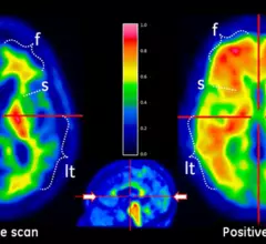
F-18 Flutemetamol (F-18 Flute) alone has been used to procure positive or negative results in visual interpretations of beta-amyloid in the brain as a tool to aid in the diagnosis and study of Alzheimer’s disease. Quantitative analysis could further supercharge this technique by bringing in more data about regional and total tracer uptake, according to a study published Aug. 21 in the Journal of Nuclear Medicine.

Novel nanoparticles pull double duty

Researchers from the University of California, Davis, have created dynamic nanoparticles that can be used to both diagnose and treat cancer.

Radiant Sage named on CIOReview list of 100 most promising technology companies

Radiant Sage LLC, a provider of on-demand clinical trial imaging infrastructure solutions, today announced that it has been named to CIOReview's list of 100 Most Promising Technology Companies. The CIOReview100 includes companies that are gaining momentum in the technology industry and making an impact.

Around the web

Positron, a New York-based nuclear imaging company, will now provide Upbeat Cardiology Solutions with advanced PET/CT systems and services. 

The nuclear imaging isotope shortage of molybdenum-99 may be over now that the sidelined reactor is restarting. ASNC's president says PET and new SPECT technologies helped cardiac imaging labs better weather the storm.

CMS has more than doubled the CCTA payment rate from $175 to $357.13. The move, expected to have a significant impact on the utilization of cardiac CT, received immediate praise from imaging specialists.